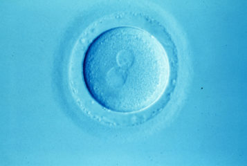
A seemingly brilliant hypothesis of PGS (preimplantation genetic screening) arose in the 1990s when Dr Yuri Verlinsky (see BioNews 519) proposed using polar body biopsies to detect chromosomal abnormalities in embryos prior to transfer in IVF. The premise behind this test was to sample the discarded 'polar bodies' released as an egg matures and is fertilised to become an embryo. The polar bodies, like the egg, should have half the full set of chromosomes and so indicate the state of the egg.
The seemingly logical assumption was that using PGS to select chromosomally healthy embryos for use in IVF would produce higher implantation, pregnancy and live birth rates.
Roughly 20 years and three generations of PGS later - its most recent one being preimplantation genetic testing for aneuploidy (PGT-A), - this add-on to IVF has as a hypothesis remained unconfirmed, and some of its expected benefits have actually been refuted.
Investigations of meiotic chromosomal defects via polar body biopsies biologically appear to make sense. Because they are technically challenging to perform they, however, never became popular. Commercial success came instead by performing biopsies on embryos on day 3 after fertilisation, at the cleavage stage, when one blastomere (sometimes two) was removed, and 6-8 chromosomes investigated with FISH (fluorescence in situ hybridisation). That moving PGS from polar bodies to cleavage stage embryos would no longer only test for meiotic but also for mitotic aneuploidies, was, however, largely dismissed as unimportant for accuracy of diagnosis.
Though never validated in predictability, sensitivity and specificity, this so-called PGS 1.0 quickly prospered. Genetic laboratories serving the IVF community, largely restricted to the testing of embryos for single gene and/or sex-linked diseases, saw an opportunity for market expansion. Though a few small prospectively randomised studies by Belgian investigators failed to demonstrate promised outcome benefits, the PGS community continued to claim outcome benefits, even after a prospectively randomised study by Dutch investigators in 2007 clearly suggested otherwise (1). This study, however, finally, set into motion an American Society for Reproductive Medicine (ASRM) review, which, later followed by other authoritative bodies, in 2008 ultimately declared PGS 1.0 ineffective (2).
As well as no benefits, the Dutch study actually suggested that, especially in older women, the procedure might negatively affect IVF outcomes (1). We had come to similar conclusions after reanalysing some of the above noted Belgian studies but had been unable to get our manuscript accepted for publication. After publication of the Dutch study, our manuscript was 'recalled' and published (3).
Following rebukes by ASRM and other professional organisations, the PGS community quickly rebooted under the argument that PGS 1.0 had failed only because of inadequate techniques and technologies. Progress in both was now possible through new more accurate diagnostic platforms: for example, moving the embryo biopsy from the cleavage stage of development to the later blastocyst stage meant more DNA was available for diagnosis as more cells (5-7) could be taken.
This new version, PGS 2.0, was again introduced to routine clinical use without prior validation studies, and quickly witnessed explosive growth.
Blastocyst-stage embryos, having undergone more cell divisions, will demonstrate more mitotic aneuploidies than cleavage-stage embryos that add to earlier meiotic aneuploidies. Embryo mosaicism – where there is a mix of chromosomal cell types, therefore, must increase between cleavage- (PGS 1.0) and blastocyst-stages (PGS 2.0). Mitotic aneuploidies have a very different clinical significance to meiotic aneuploidies: mouse and human stem cell experiments suggest that embryos possess an innate ability to self-correct these chromosomal abnormalities. Therefore, they denote a different risk profile for human embryos than meiotic aneuploidies.
All of this points toward increasing risks for false-positive diagnoses by moving biopsies from cleavage stage to blastocyst-stage. This was, however, again dismissed by the PGS community as insignificant for accuracy.
PGS 2.0 considered embryos with any amount of chromosomally abnormal DNA as 'aneuploid' and, therefore, unsuitable for transfer. This leads to the possibility that large numbers of potentially chromosomally normal embryos were erroneously discarded. In 2012, our IVF centre under experimental protocol, therefore, started offering patients who had no 'euploid' (chromosomally normal) embryos in IVF cycles, transfers of selected aneuploid embryos. These embryos had selected monosomies – with only one chromosome of a pair present, or later by 2014 this was expanded to selected trisomies – where there is an additional chromosome to a pair.
We reported the first five chromosomally healthy births from such transfers in October 2015 (4), shortly followed by similar results from Italian colleagues (5). By now, over 100 healthy pregnancies and deliveries have been reported worldwide following such transfers.
Considering these reports and blatant discrepancies in PGS results between prominent PGS laboratories, but also between multiple biopsies from same embryos in same laboratories (6), by July 2016, PGS 2.0 was no longer defensible. The Preimplantation Genetic Diagnosis International Society (PGDIS) published new PGS guidelines, which radically changed practice to PGT-A.
Based on a so-called 'threshold concept', PGS laboratories from this point on moved from the reporting of euploid/aneuploid to the reporting of euploid, mosaic and aneuploid. The diagnosis now depended on how much 'aneuploid' DNA a single biopsy specimen contained: with up to 20 percent DNA, embryos were considered 'normal-euploid'; between 20-80 percent 'mosaic'; and only if the aneuploid DNA load was above 80 percent, embryos were considered 'aneuploid'.
The PGDIS, however, offered no validation studies for these new definitions. Indeed, the 20 percent cut-off between euploid and mosaic was, likely, only a technical 'accident' because with currently available technology Next Generation Sequencing (NGS) is only able to detect mosaicism above 20 percent DNA load.
The new PGDIS guidelines also permit selected transfers of mosaic embryos. The irrationality of those guidelines is, however, demonstrated by an embryo with 19 percent mosaicism now being allowed to be transferred as 'normal'; yet an embryo with 21 per cent aneuploidy load is considered 'abnormal-mosaic' and not transferred.
Even the most current diagnostic methods utilised by the PGS industry make little sense biologically: detection of mitotic (in contrast to meiotic) aneuploidies depends on where in the embryo a biopsy is taken from. If the trophectoderm (the outer layer of cells in the embryo) is overwhelmingly euploid and, by accident, a biopsy is taken from a small aneuploid clone, resulting in all 5-7 cells of the biopsy being aneuploidy, the embryo will under new PGDIS guidelines be declared aneuploidy - though very likely a false-positive diagnosis. The reverse can also happen to give a false-negative diagnosis.
In both situations, the embryo is, however, really mosaic yet not recognised as such under the new PGDIS guidelines. These define an embryo as mosaic only if, again by accident, a borderline area between normal and abnormal cells is biopsied, resulting in a mix of euploid and aneuploid cells. PGDIS guidelines thereby greatly underestimate the true prevalence of trophectoderm mosaicism.
In other words, a single 5-7-cell trophectoderm biopsy is mathematically incapable of accurately determining the true status of the complete trophectoderm (9). Moreover, the trophectoderm - from which the placenta arises, does not always fully reflect the inner cell mass, from which the fetus arises. As has been known for decades, aneuploid clones are quite common in mature placentas of perfectly normal pregnancies with euploid offspring.
In summary, three iterations of PGS have failed in improving IVF outcomes - the basic motivation of the PGS hypothesis. More likely is that as a consequence of discarding good quality embryos because of frequent false-positive diagnoses, the procedure in poorer prognosis patients appears to reduce pregnancy and live-birth chances. Paulson recently calculated this risk at about 40 percent (10). We believe that it is even higher.
The paradox, however, is that the PGS community has for over 20 years constantly presented new excuses for the lack of promised outcomes, and persistently moved goal posts and demanded that critics prove their point, when basic rules of medical evidence mandate that the burden of proof lies with proponents of new treatments. After two decades of clinically unproven utilization of PGS, the erroneous disposal of thousands of perfectly normal embryos, it is time for proponents of PGS/PT-A to finally prove, beyond reasonable doubt, that the procedure does indeed have clinical value. Until then, PGS 3.0 should not be offered as routine add-on to IVF, except in such validation studies and under experimental consents.
1) Mastenbroek S et al. In vitro fertilization with preimplantation genetic screening N Engl J Med. 2007 Jul 5;357(1):9-17.
2) Practice Committee of the American Society for Reproductive Medicine, Preimplantation genetic testing: a Practice Committee opinion. Fertil Steril 2008; 90:S136-143
3) Gleicher N, Weghofer A, Barad D. Preimplantation genetic screening, 'established' and ready for prime time? Fertil Steril 2008;89(4):780-788
4) Gleicher N, Vidali A, Braverman J, Kushnir VA, Albertini DF, Barad DH., 2015. Further evidence against use of PGS in poor prognosis patients: report of normal births after transfer of embryos reported as aneuploid. Fertil Steril 2015;104(Suppl) 3:e9
5) Greco E, Giulia Minasi M, Florentino F., Healthy babies after intrauterine transfer of mosaic aneuploid blastocysts. N Engl J Med 2015; 373:2989-2090
6) Gleicher N, Vidali A, Braverman J, Kushnir VA, Barad DH., Hudson C, Wu YG, Wang Q, Zhang L, Albertini DF. Accuracy of preimplantation genetic screening (PGS) is compromised by degree of mosaicism of human embryos. Reprod Biol Endocrinol 2016;14:54
7) Munné S, Blazek J, Large M, Martinez-Ortiz PA, Nisson H, Liu E, Tarozzi N, Borini A, Becker A, Zhang J, Maxwell S, Grifo J, Barbariya D, Wells D, Fragouli E. Detailed investigation into the cytogenic constitution and pregnancy outcome of replacing mosaic blastocysts detected with the use of high-resolution next-generation sequencing. Fertil Steril 2017; http://dx.doi.org/10.1016/j.fernstert. 2017.05.002
8) Kushnir VA, Darmon S, Albertini DF, Barad DH, Gleicher N. Degree of mosaicism in trophectoderm does not predict pregnancy potential: a corrected analysis of pregnancy outcomes following transfer of mosaic embryos. Submitted for publication;
9) Gleicher N, Metzger J, Croft G, Kushnir VA, Albertini DF, Barad DH., 2017. A single trophectoderm biopsy at blastocyst stage is mathematically unable to determine embryo ploidy accurately enough for clinical use. Reprod Biol Endocrinol 2017;15(1)23
10) Paulson RJ. Preimplantation genetic screening: what is the clinical efficiency? Fertil Steril 2017;108(2):228-230


Leave a Reply
You must be logged in to post a comment.