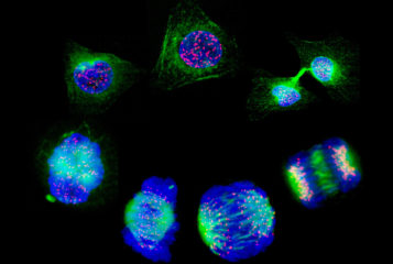
Image by NIMR/Francis Crick Institute via the Wellcome Collection. Depicts cultured cells stained to show the nucleus in blue, the tubulin in green and the mitochondria in red.
382 articles
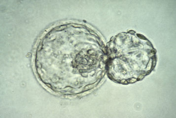
Image by K Hardy via the Wellcome Collection. Depicts a human embryo at the blastocyst stage (about six days after fertilisation) 'hatching' out of the zona pellucida.
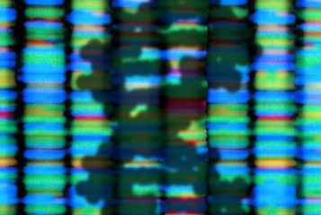
Image by Peter Artymiuk via the Wellcome Collection. Depicts the shadow of a DNA double helix, on a background that shows the fluorescent banding of the output from a DNA sequencing machine.
Image by Peter Artymiuk via the Wellcome Collection. Depicts the shadow of a DNA double helix, on a background that shows the fluorescent banding of the output from a DNA sequencing machine.
Image by K Hardy via the Wellcome Collection. Depicts a human embryo at the blastocyst stage (about six days after fertilisation) 'hatching' out of the zona pellucida.
Image by Peter Artymiuk via the Wellcome Collection. Depicts the shadow of a DNA double helix, on a background that shows the fluorescent banding of the output from a DNA sequencing machine.
Subscribe to BioNews and other PET updates for free
BioNews, published by the Progress Educational Trust (PET), provides news and comment on genetics, assisted conception, embryo/stem cell research and related areas.