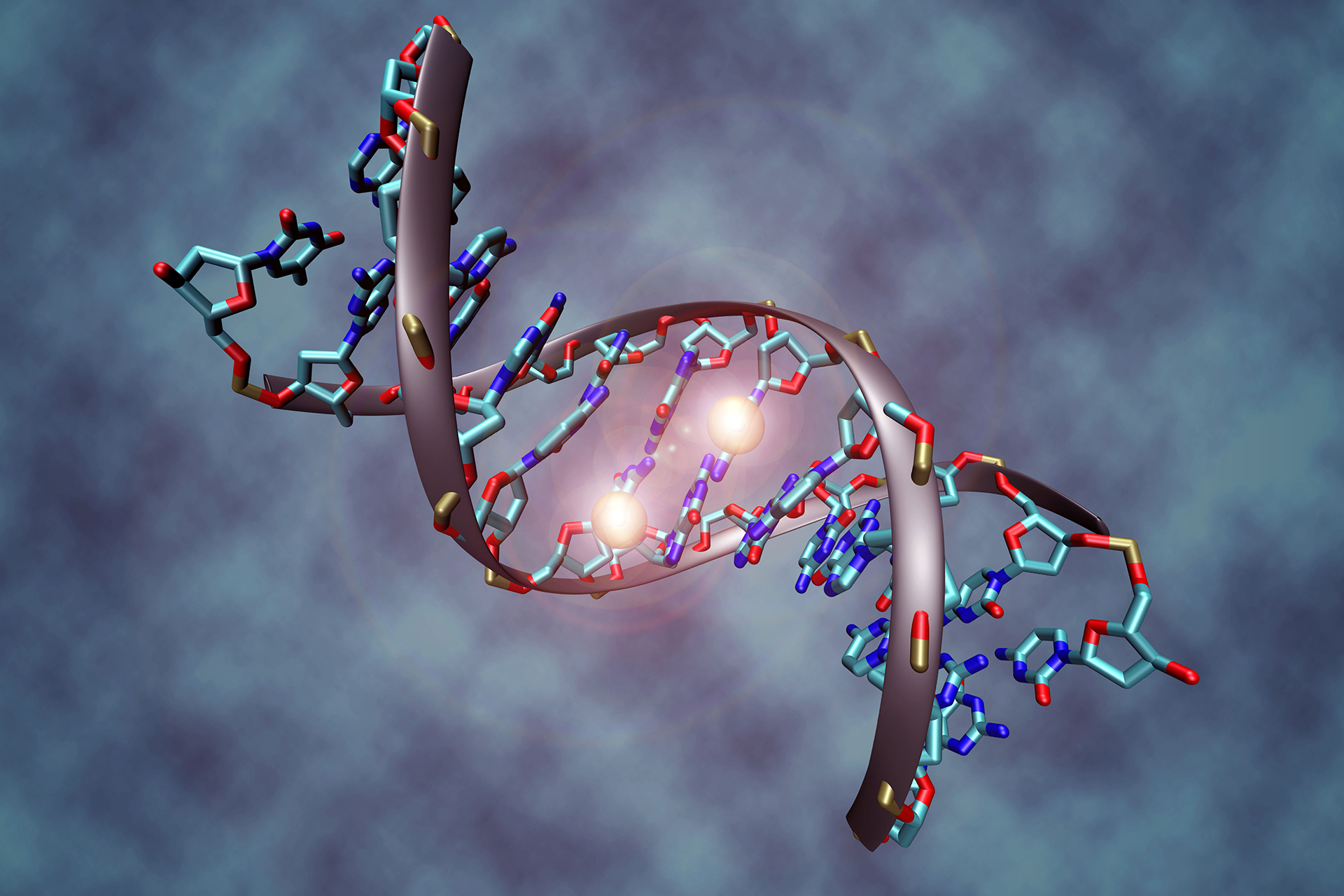
The three-dimensional structure of the X chromosome and how it is silenced in female cells has been demonstrated by a new computational method.
Using artificial intelligence, US scientists have been able to probe the 3D structure of the X chromosome and investigate how RNA causes the chromosome to fold and unfold in female embryos, leading to X inactivation.
'This is the first time we've been able to model all the RNA spreading around the chromosome and shutting it down,' said lead author Dr Anna Lappala from the Los Alamos National Laboratory in New Mexico. 'From experimental data alone, which is 2D and static, you don't have the resolution to see a whole chromosome at this level of detail. With this modelling, we can see the processes regulating gene expression.'
As female mammalian embryos are conceived with two X chromosomes, one is 'switched off' in each cell. X inactivation occurs when noncoding RNA found on the X chromosome engulfs the chromosome and recruits numerous protein complexes to inactivate and silence it.
To investigate X inactivation, researchers used experimental data from Harvard Medical School in Boston and Massachusetts General Hospital to develop the 4DHiC method that runs on supercomputers at the Los Alamos National Laboratory. 4DHiC extract 3D information from experimental mouse genomic data and use X inactivation as a model to examine the time evolution of 3D chromosome architecture during changes in gene expression.
The method was able to reveal in greater detail the folding and unfolding process of the X chromosome as it transitions from an active to inactive form. In a visualisation created by the researchers to show this process, RNA particles are shown to engulf and penetrate the inner structure of the chromosome to target and bind to the X chromosome inactivation centre to silence the chromosome.
The Los Alamos team are now developing a browser where scientists can upload genomic data to view chromosomes in 3D at different magnifications. They hope this new method will provide a greater understanding of gene expression and may reveal new treatments for genetic conditions.
'What's been missing in the field is some way for a user who's not computationally savvy to go interactively into a chromosome,' said Professor Jeannie Lee, professor of genetics at Harvard Medical School. She compared using the 4DHiC method to Google Earth, where 'you can zoom into any location on an X chromosome, pick your favourite gene, see the other genes around it, and see how they interact.'
The study was published in Proceedings of the National Academies of Science.
Sources and References
-
Supercomputers reveal how X chromosomes fold, deactivate
-
New 4D visualization shows epigenetic processes unfold over time
-
Supercomputer reveals how the X chromosome folds and deactivates
-
3D models reveal hidden process in X chromosome inactivation
-
Four-dimensional chromosome reconstruction elucidates the spatiotemporal reorganization of the mammalian X chromosome


Leave a Reply
You must be logged in to post a comment.