As you start to walk out on the way, the way appears. (Rumi)
Almost a century ago, Professor Hans Spemann, a German biologist from the University of Freiburg, and his PhD student Hilde Mangold defined a group of cells in newt embryos that triggered development of the central nervous system. They transplanted that group of cells from one site in the embryo to another using tiny microsurgical tools that they had developed, made from glass and hair. The transplanted cells induced development of the central nervous system in the new place in the embryo too.
Other researchers confirmed the finding and the group of the cells with that special property was named the 'Spemann-Mangold organiser'. In 1935, Professor Spemann received the Nobel prize for medicine for this work.
The molecular mechanisms behind this process were deciphered decades later. Cells in organisers were found to secrete proteins that influence the pattern of embryo development.
Similar structures with a role in body patterning were later detected in fish (the embryonic shield), birds (Hensen's node) and rodents. However, for nearly 100 years, the organiser remained elusive in primates, including humans.
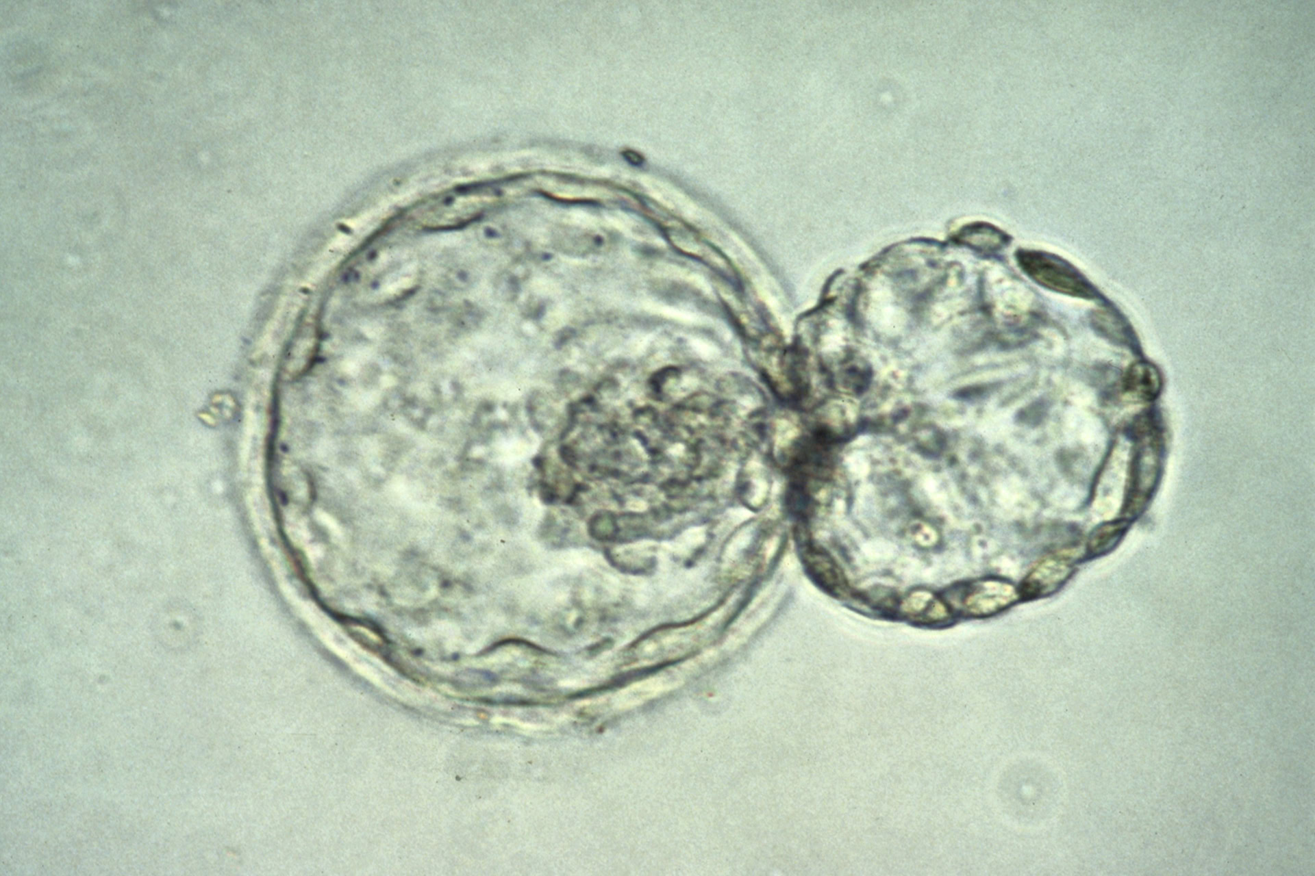
The organiser has now been seen for the first time in human-chicken chimera embryos (see BioNews 951). This development discovery was made possible by developing a very specific micropattern technology for growing human embryonic stem cells on.
The stem cell biologist Dr Ali Brivanlou and physicist Dr Eric Siggia from Rockefeller University in New York City joined forces to create this micropatterned surface technology. The growing surface enabled human embryonic stem cells (hESCs) to self-organise. The surface allowed the cells grow and generate the geometry seen in early embryonic development in vivo. The cells, confined to disc-shaped colonies less than a millimetre in size, were stimulated with specific growth factors to form self-organised differentiation patterns in concentric radial patterns.
The system worked really well for mimicking the self-organising abilities of in vitro attached human embryos. It has provided us with a new understanding of early human embryonic development beyond the blastocyst stage. Over a period of a few days, the cells formed patterns reminiscent of embryos going through gastrulation, the stage at which they develop from a blob of cells to a structure with a defined front, back, top and bottom (see BioNews 932).
Single-cell time-lapse imaging and other customised tools can be used to see in finer detail the ongoing cell-to-cell signalling and relationship between the cells that will form distinct layers in the embryo.
In a recent publication, using this quite sophisticated micropattern culture system, Dr Brivanlou and Dr Siggia demonstrated that the organiser does exist in humans. In mouse embryos, patterning of the gastrulation stage embryo is governed by specific growth factors and small molecule inhibitors, so they hypothesised that this might be a case in humans too.
Micropatterned colonies were exposed to various combinations of these factors and inhibitors. And indeed, they were able to generate patch of cells that secreted factors known to be produced by organisers and its derivatives in other systems.
So far, all the evidence supported the hypothesis that humans had the structure. However, the ultimate proof of a working organiser, as defined by the classic amphibian experiments, is that this particular group of cells can induce a secondary axis (a different body plan) – when grafted onto embryos.
In a beautifully done experiment, Dr Brivanlou and Dr Siggia did just that. Micropatterned stem cell colonies were treated with a particular combination of growth factors that would induce formation of organiser. Next, they were grafted onto a chick embryo where they directly contributed to the secondary axis. They continued to differentiate in their new environment, contributing to a secondary axis.
These experiments closed the loop opened nearly 100 years ago, demonstrating that the embryo's organiser structure is conserved from frogs to humans.
What is the next step for this biologist-physicist tandem? Dr Brivanlou and Dr Siggia have founded a company in San Francisco called Rumi Scientific, named after the 13th century Persian Sunni Muslim poet, jurist, Islamic scholar, theologian and Sufi mystic, Rumi. Their goal for the company is to use this technology to develop a process for screening and testing for genetic diseases.
Their first chosen target is Huntington's disease. They have now demonstrated that human embryonic stem cells with the Huntington's disease mutation leave a unique signature on their micropatterned colonies, easily distinguishable from mutation-free counterparts. Furthermore, they have also identified compounds capable of reverting the Huntington's signature back to normal.


Leave a Reply
You must be logged in to post a comment.