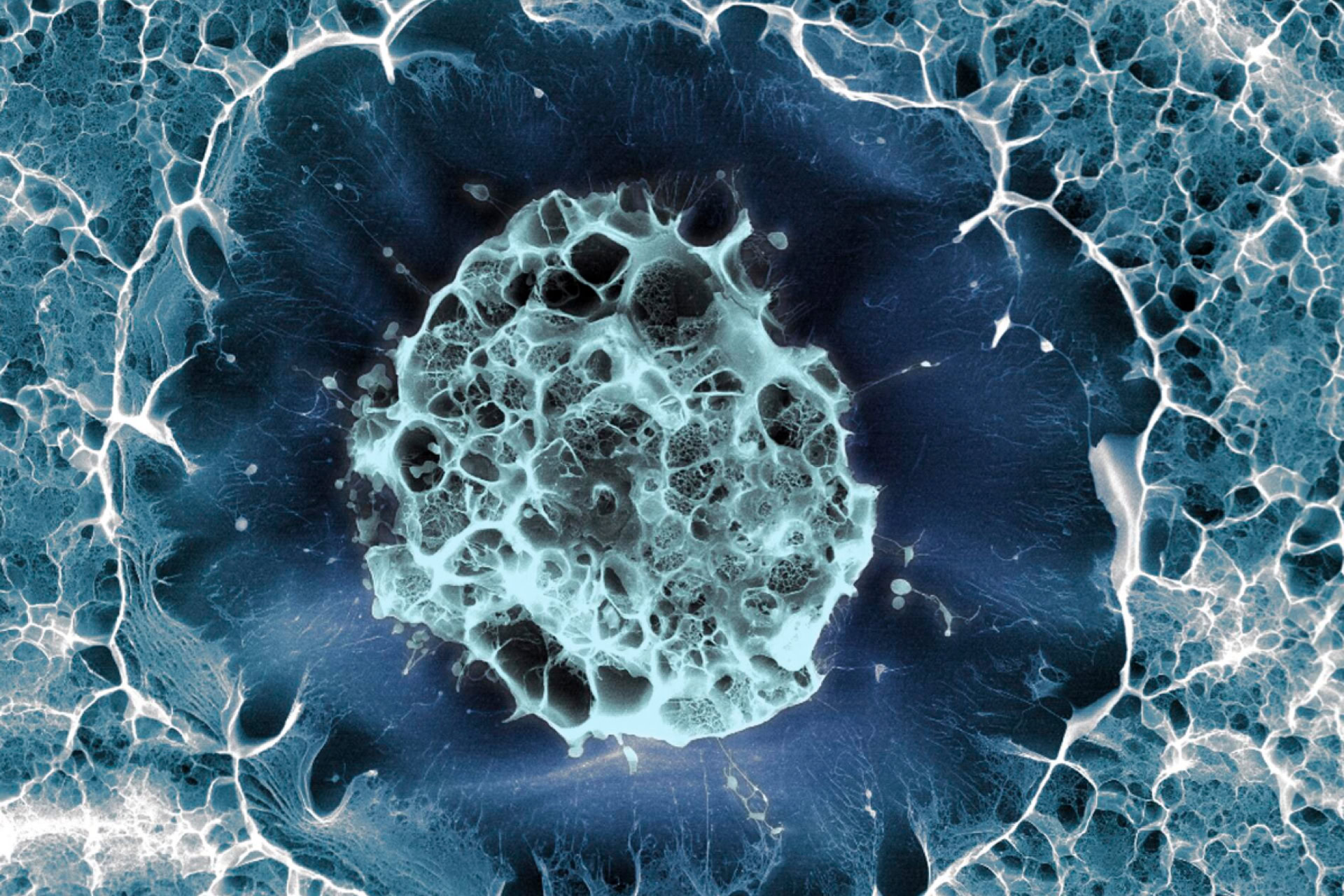
Researchers have successfully filmed the division of stem cells in an adult mouse brain, for the first time.
The study has shown how the hippocampus, a brain structure involved in learning and memory, generates new cells throughout life. This may help lead to a deeper understanding of the role of cell renewal in the brain.
'In the past, it was deemed technically impossible to follow single cell stem cells in the brain over time given the deep localisation of the hippocampus in the brain,' said senior author Professor Sebastian Jessberger, from the Brain Research Institute at the University of Zurich, Switzerland.
The researchers used a microscopy technique called two-photon imaging to capture pictures of the stem cells dividing. They first removed the outer layers of brain tissue in living mice to uncover the hippocampus and genetically labelled 63 individual stem cells. Pictures were taken every 12 to 24 hours for two months to track the development and maturation of the new neural cells.
Most of the stem cells had only a few rounds of cell division before they matured into neurons, which integrated into the adult mouse hippocampus and no longer divided. The short maturation process may explain why the number of new neurons decreases dramatically with age.
The new study, published in Science, was made possible due to collaborations with experts in deep-brain imaging and theoretical stem cell modelling, from the Brain Research Institute and the University of Cambridge, UK.
As well as deepening our understanding of how brain cells develop throughout life, it is hoped the study will help advance research into therapies for human diseases.
'In the future, we hope that we will be able to use neural stem cells for brain repair – for example for diseases such as cognitive ageing, Parkinson's and Alzheimer's disease or major depression,' Professor Jessberger concluded.


Leave a Reply
You must be logged in to post a comment.