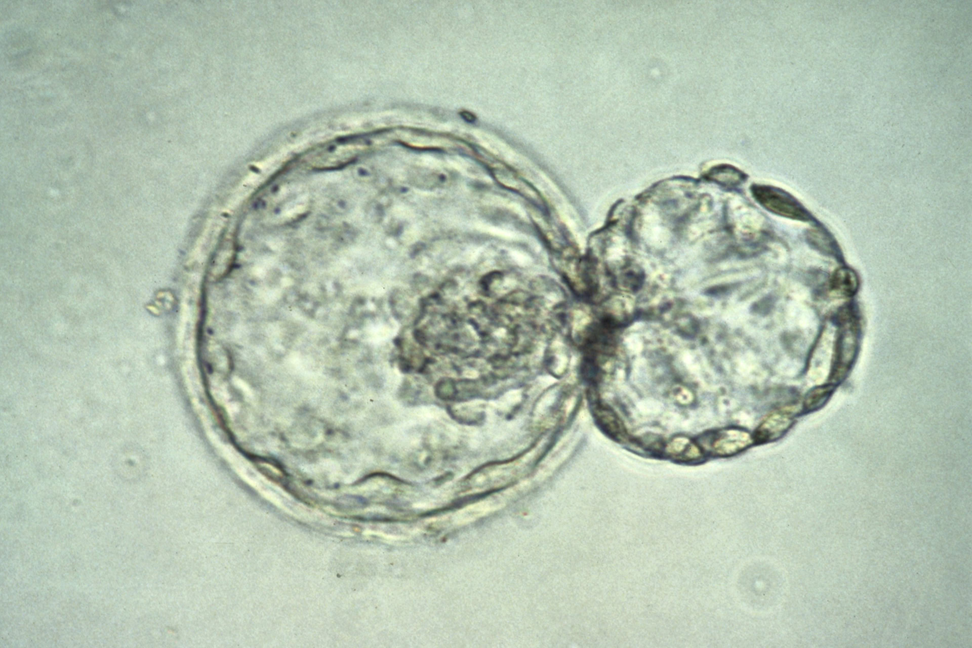
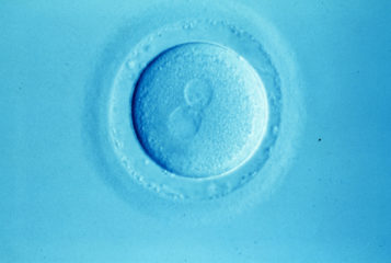
An interactive map exhibiting the development of the early human embryo in unprecedented detail has been created by a team of Dutch researchers.
The atlas, which shows embryonic development from conception to two months, offers the opportunity to explore how the fetus as a whole matures, in addition to the individual systems that constitute the body, such as the cardiovascular system.
Bernadette de Bakker, co-lead on the project, which was based at the University of Amsterdam, said that the map 'is the first to present such a large amount of data based on such a large number of human embryos. With this atlas and database we are able to quantify very precisely embryonic growth and the growth and position of specific organs'.
The research, published in Science, involved analysing around 15,000 histological sections obtained from the Carnegie Collection of human embryos at the National Museum of Health and Medicine in Washington DC, which were digitised to produce accurate, interactive 3D models. Fourteen models span over the first nine-and-a-half weeks of development, allowing the user to learn about 150 organs and structures within each model and monitor changes in the embryo over time.
Magdalena Zernicka-Goetz, professor of mammalian development at the University of Cambridge, who was not involved in the research, told The Guardian that 'the dearth of knowledge [in human embryology] is down to barriers in experimental study', such as the difficulty in obtaining embryonic samples, and the limit of 14 days for live human embryo culture.
De Bakker said the current knowledge of human development is predominantly based on findings obtained from specimens examined in the early 19th century.
'Everyone thinks we already know this, but I believe we know more about the moon than about our own development,' she said, adding that the atlas highlights significant differences between animal models, such as the frequently used mice and chick embryos, and actual human embryo development.
'We discovered that some organs in humans develop earlier than they first arrive in chick or mouse embryos, and some [develop far] later,' De Bakker said. This may have consequences for developmental toxicology when testing new drugs and chemicals, for example.
Additionally, De Bakker said the work will improve scientists' general understanding of early development and allow researchers to explore developmental diseases in greater detail, as they can see where and how embryo maturation can go wrong: 'It is important to understand normal human embryology to clarify how inborn defects and congenital malformations occur.'
Sources and References
-
3-D embryo model shows early human development in astonishing details
-
3D embryo atlas reveals human development in unprecedented detail
-
An interactive three-dimensional digital atlas and quantitative database of human development
-
Detailed human embryo development revealed in three-dimensional 'atlas'


Leave a Reply
You must be logged in to post a comment.