
A technique for monitoring embryo health could increase the chance of IVF couples having a healthy baby, according to a study from researchers at a private fertility clinic.
The method involves taking thousands of pictures of embryos to track their development before they are implanted in the womb. The clinic, one of the few in the UK to offer this technique, claims that this can boost IVF success by more than 50 percent.
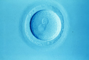
IVF is extremely costly, and typically has a low chance of success. One of the most common causes of failed IVF is aneuploidy, when implanted embryos have too many or too few chromosomes. Currently, the only way to check an embryo for chromosomal abnormalities is to remove a cell, which then undergoes genetic screening. This procedure costs about £2,500, and the act of removing a cell can cause developmental problems further down the line.
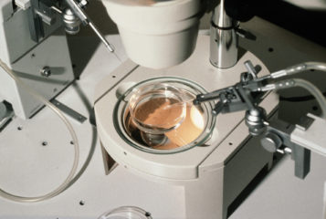
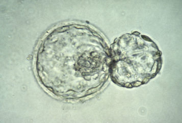
Now, scientists at CARE fertility clinic in Nottingham, UK, have developed an alternative approach based on time-lapse imaging. Fertilised eggs are placed in an incubator, which is hooked up to a video system capable of capturing thousands of images documenting the embryos' development from conception to the blastocyst stage (70 to 100 cells). 'This is almost like having the embryo in the womb with a camera on them', explains CARE's managing director Dr Simon Fishel.
Studying these images reveals certain trends. The team found, for example, that abnormal embryos took around six hours longer to form a blastocyst than healthy ones. They came up with an algorithm for predicting embryonic health, and the likelihood that a particular embryo would lead to a successful birth.
'In the 35 years I have been in this field this is probably the most exciting and significant development that can be of value to all patients seeking IVF', Dr Fishel told the BBC. The clinic claims the new technique could increase the success rate for IVF treatment to 78 percent - over three times the UK national average.
They investigated 88 embryos from 69 couples visiting the clinic, using the time-lapse technology to classify embryos as having high, medium or low risk of chromosomal defects. Eleven babies were born from the low risk group (61 percent success rate) and five from the medium risk group (19 percent success rate). No embryos from the high-risk group successfully implanted.
Only about a dozen IVF clinics in the UK offer the £750 imaging service, but CARE researchers say that if the technique were to be adopted by the NHS, it could boost success rates and reduce the costs of IVF.
But others are more cautious. Dr Allan Pacey, chair of the British Fertility Society, said: 'All too often developments in IVF are trumpeted as advances when they remain unproven. In this case, whilst this is a good piece of science, before we splash this on the front page it should be subject to full randomised control trials'.
The study was published in the journal Reproductive BioMedicine Online.
Sources and References
-
Retrospective analysis of outcomes after IVF using an aneuploidy risk model derived from time-lapse imaging without PGS
-
IVF 'may be boosted by time-lapse embryo imaging'
-
IVF time-lapse study needs more evidence
-
Time-lapse spots faulty embryos before IVF
-
IVF could be revolutionised by new technique, says clinic
Related Articles
How fast is too fast? Innovation in IVF and the burden of proof
The speed of how technological innovations are deployed in infertility care is discussed here by Dr Rita Vassena...
Artificial intelligence learns to identify good embryos for IVF
Researchers have trained artificial intelligence software to grade human embryos with a high degree of accuracy...
New imaging technique could help pick embryos for IVF
A new imaging technique can help assess the quality of early-stage embryos...
3D human embryo development 'atlas' created
An interactive map exhibiting the development of the early human embryo in unprecedented detail has been created by a team of Dutch researchers...
First study of chromosome test suggests increase in IVF success
An IVF test which checks whether embryos carry the correct number of chromosomes could improve the chances of a successful pregnancy, a clinical trial suggests. The test — developed by the biotech company Blue Gnome — is used five days after an egg has been fertilised and helps doctors select which embryos should be implanted during IVF treatment...
Progress Educational Trust Conference: Should Assisted Conception Always Be Evidence-Based?
In what is now synonymous with Progress Educational Trust (PET)'s ethos, the final session of the annual conference, 'The Best Possible Start in Life: The Robust and Responsive Embryo', was a free-form debate. Following on from the previous sessions where a wealth of eminent researchers gave informative and often provocative talks, Guardian columnist Zoe Williams had the task of chairing what proved to be an entertaining debate...
Embryo's survival can be predicted based on egg's movement
Rhythmic activity detected in newly fertilised mouse eggs may provide a novel and non-invasive screening method for identifying embryos most likely to survive a full-term pregnancy, according to research published in Nature Communications...
Imaging IVF embryos can predict survival
US researchers have developed a means to predict which human embryos produced through IVF are most likely to result in healthy births. Researchers filmed 242 one-cell embryos and predicted, with more than 93 percent accuracy, those that would survive up to five days. These findings may improve the success rate of IVF....
Leave a Reply
You must be logged in to post a comment.