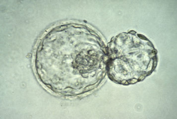
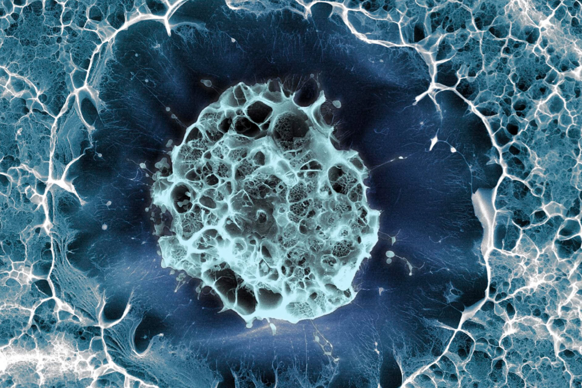
The first 'virtual embryo' has been created to help understand early embryo development and the evolution of a single cell into many cells of different types.
Publishing their results in the journal Cell, researchers from the European Molecular Biology Laboratory (EMBL) in Heidelberg, Germany, and from the University of Padua School of Medicine, Italy, have described the development of every single cell in the embryo.
'How are the many different cell types in the body generated during embryonic development from an egg, which is only a single cell? This is one of the most fundamental questions in biology,' said Dr Pierre Neveu, group leader at EMBL, explaining the rationale behind the study.
All organisms begin as a single cell, which divides, producing new cell lines predetermined to become various tissues and structures. A representation of cell lineages in embryo development, accounting for every cell in space and time, has not been achieved until now.
'So far we have lacked a comprehensive understanding of the gene expression programmes. These instruct individual cells to form the different cell types necessary to build an embryo,' explained first author Dr Hanna Sladitschek.
The researchers modelled the virtual embryo on a marine organism called Phallusia mammillata. Each individual organism has the same number of cells, which makes it easier to reconstruct its embryonic development by combining observations from many samples.
To generate the virtual embryo, the team used high-resolution single-cell transcriptome. The transcriptome is all the RNA molecules in a cell, providing a snapshot of which genes are switched on or off in that cell at a given time.
This was combined with data from microscopic observation of the structures formed by P. mammillata dividing cells, to generate a detailed four-dimensional (4D) atlas of embryonic development from one cell to 64 cells.
Furthermore, by using high-level single-cell RNA sequencing, the team designed a computational framework, MorphoSeq, which categorised the embryos into cell types, reconstructed directly from the physical position and lineage history of each cell. From there, the cells were linked to the high-resolution 4D imaging data.
After the first seven cell divisions, the nerve cord, brain, germ cells, blood cell precursors, and muscles are already determined. This study presents the first full description of early development accounting for every single cell in an embryo.
'Our model shows that it is possible to know the location and history of an individual cell by analysing its gene expression,' said Dr Neveu. 'In addition, we find that while the regulation of gene expression is very precise within an embryo, differences in developmental timing explain the observed variation between individual embryos.'
The researchers hope that the MorphoSeq framework can be readily adapted to mammals' embryonic development.
'Our studies represent a leap forward in the emerging field of developmental genomics,' said co-author Dr Lars Hufnagel, from the University of Padua School of Medicine. 'Now that we have worked with an organism with a small number of cells, it will, of course, be very interesting to extend our work to mammals, which have many more cells!'
Sources and References
Related Articles
Genetic and epigenetic causes of IVF embryo arrest discovered
Changes that occur to DNA that could cause IVF embryos to stop dividing have been identified...
Early pregnancy loss may be explained by embryo development discovery
Key molecular events regulating early embryo development have been revealed for the first time...
Organoids mimic the early development of the heart in mouse embryos
Organoids can be used to study early stages of heart development in mouse embryos, scientists say...
Embryo development 'clock' differs in mouse and human
A newly discovered mechanism explains why pregnancies last longer in some species than others...
Human stem cells used to mimic early embryo development
Using human stem cells, scientists from the University of Cambridge have developed a model for early embryo development...
Mouse artificial 'embryo' created from stem cells
For the first time, an artificially create a mouse embryo has successfully passed a critical developmental milestone in the lab...
Leave a Reply
You must be logged in to post a comment.