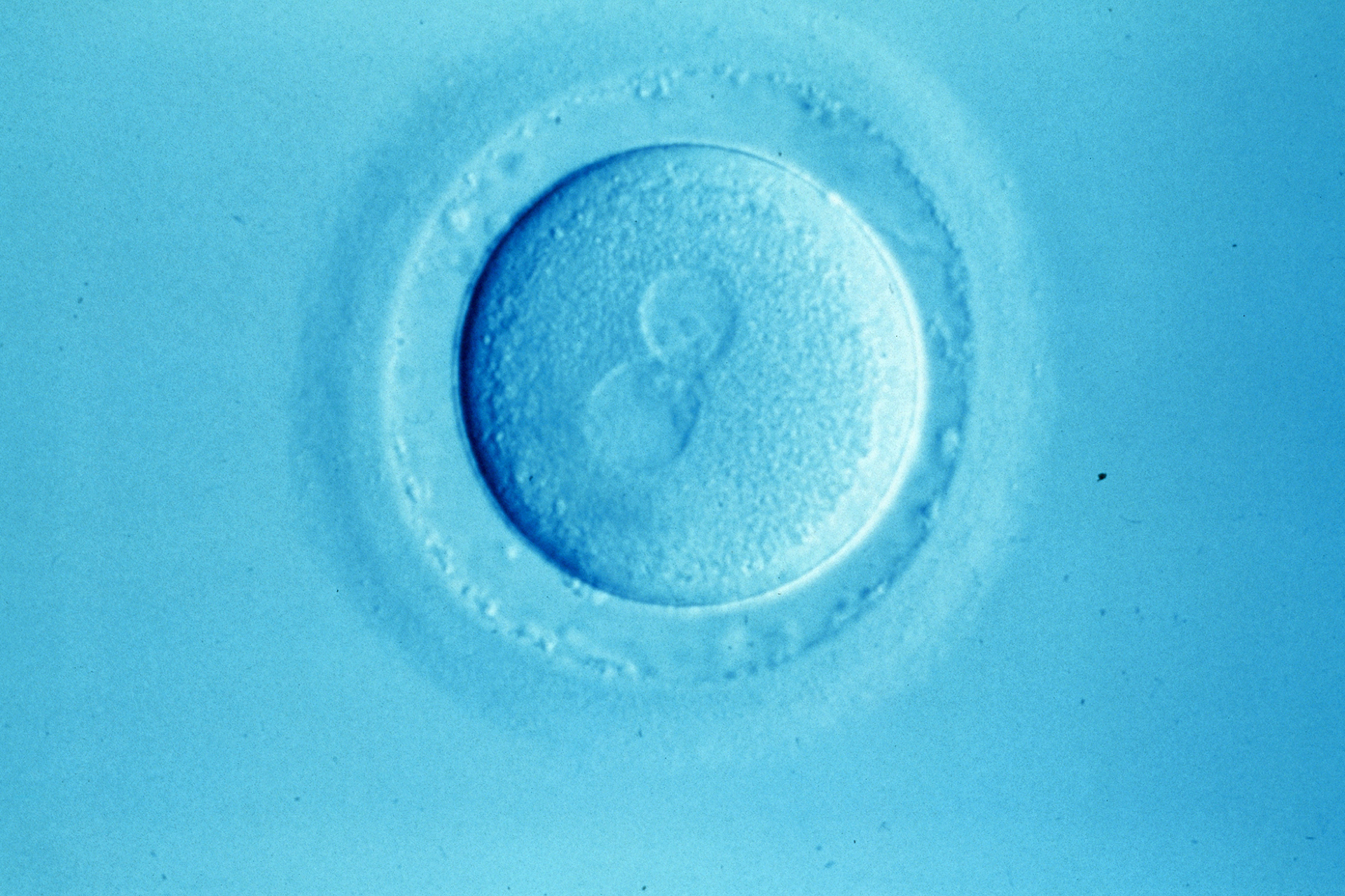
The US Food and Drug Administration has approved a time-lapse imaging tool designed to improve the embryo selection in IVF for use in the USA.
The Early Embryo Viability Assessment, or 'Eeva', developed by Auxogyn, uses proprietary software to image the development of embryos in the laboratory. The company says this information can help doctors to determine which ones are most likely to be viable.
A clinical trial showed that using Eeva along with morphology techniques meant that the odds of an embryo reaching blastocyst stage were 2.57 times greater than those not predicted to reach blastocyst stage, around five to six days. The company said the results showed a 53 percent increase in odds ratio over traditional morphology techniques alone.
Dr Michael Glassner, one of the doctors running the trial and division head of infertility at Bryn Mawr Hospital, Pennsylvania, said: '[Eeva] is helpful beyond words. It's going to give more clarity to the patient. It's going to give a higher pregnancy rate. The miscarriage rate goes down. It's just going to change the field'.
The method behind the test was developed by researchers at Stanford University in 2010. It relies on imaging the first few cell divisions of developing embryos and timing how long these take to happen, with the time between each cell division being a good prediction of embryo viability. Auxogyn acquired a license to develop products based on this method shortly after the work was published, and patented the technique in 2012.
The method was approved for use in the EU in 2012 and is also licensed for use in Canada. Auxogyn plans to commercialise the test in the USA later this year. 'We're excited to receive the de novo FDA clearance for the Eeva System and believe this marks a significant milestone in the field of IVF', said Lissa Goldenstein, CEO of Auxogyn.
'At Auxogyn, we are committed to improving the IVF journey for couples and giving them a better chance of a successful outcome'.


Leave a Reply
You must be logged in to post a comment.