Artificial heart ventricles that closely resemble the structure of the human heart have been developed by researchers at Harvard University, Boston, Massachusetts.
A new tissue engineering technique was used to spin fibres into a helical structure on which human stem cell derived cardiomyocytes (heart muscle cells) were grown. Ventricles developed using this technique were able to eject more liquid compared to those with circumferentially aligned cells. Published in Science, this research aids in the development of hearts for transplantation as well as providing a model to research heart disease.
'Since 2003, our group has worked to understand the structure-function relationships of the heart and how disease pathologically compromises these relationships,' said senior author Professor Kevin Kit Parker of Harvard University. 'This work is a major step forward for organ biofabrication and brings us closer to our ultimate goal of building a human heart for transplant.'
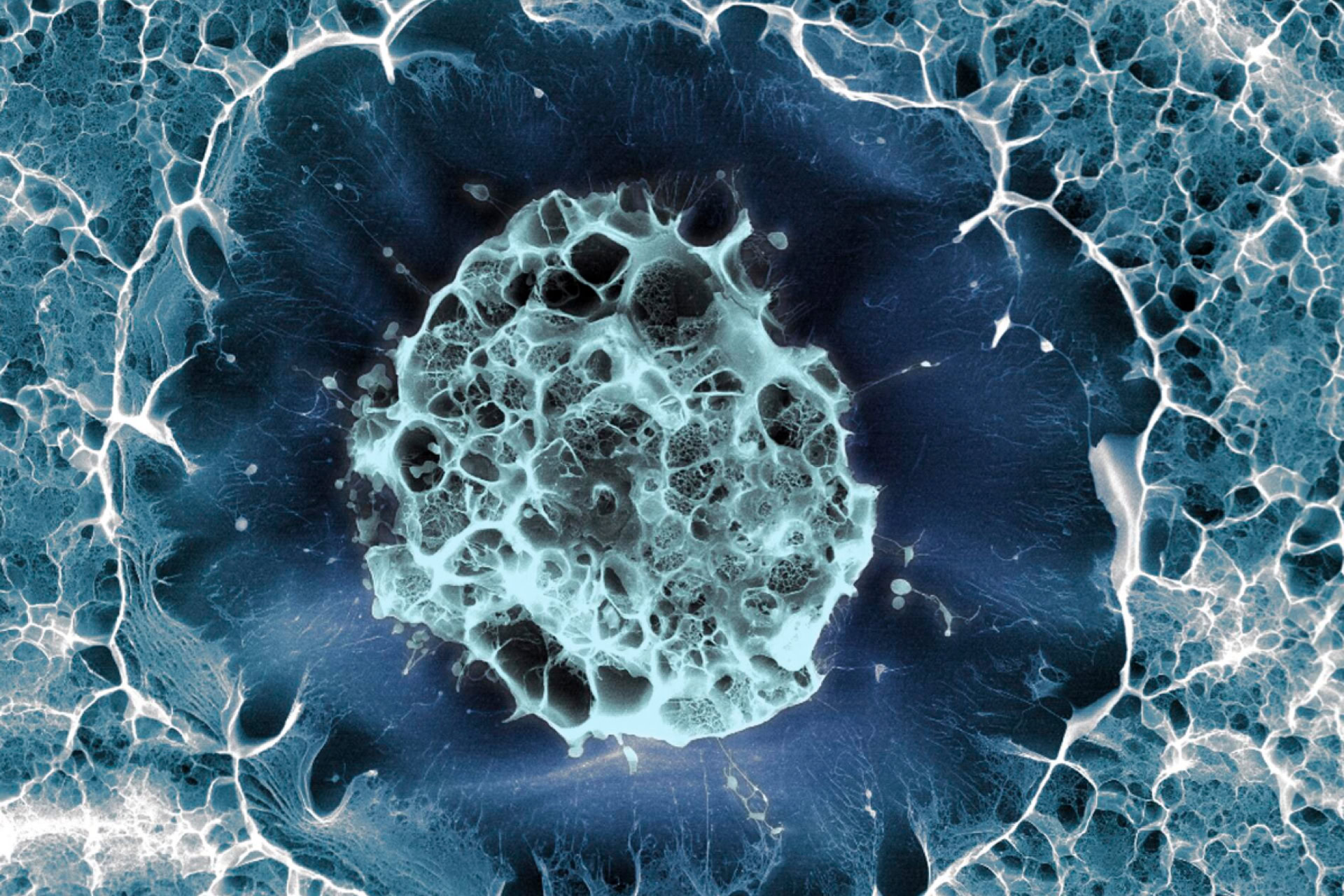
The researchers used a new method of manufacturing tissue engineering scaffolds called focused rotary jet spinning (FRJS). This technique works a bit like a candy floss machine, spinning a liquid polymer solution through tiny holes onto a collector. By angling and rotating the collector, the team were able to create helical scaffolds at the nano- and micrometre scale.
Human stem cell derived cardiomyocytes were seeded onto the scaffolds and within a week had grown thin layers of beating tissue following the helical alignment. These ventricles were able to beat in a wringing motion, mimicking that of the human heart.
'The human heart actually has multiple layers of helically aligned muscles with different angles of alignment. With FRJS, we can recreate those complex structures in a really precise way, forming single and even four chambered ventricle structures,' said Dr Huibin Chang of Harvard University, co-first author of the paper.
Ventricles grown using helical alignment performed better than circumferentially grown ventricles on ventricle deformation, speed of electrical signalling and fraction ejection. The team also demonstrated that this technique could be scaled up to the size of a human heart and even to the size of a minke whale heart.
The spiral-like arrangement of heart muscle was first noted in 1669. Centuries later, in 1969, Professor Edward Sallin theorised that this arrangement is critical to the expulsion capabilities of the human heart. However, studying this architecture has proven difficult. Now, decades later, this team have been able test Professor Sallin's theory and answer questions that are centuries old.
'Our goal was to build a model where we could test [Professor] Sallin's hypothesis and study the relative importance of the heart's helical structure,' said Dr John Zimmerman of Harvard University, co-first author of the paper.
Sources and References
Related Articles
Donor stem cells offer hope for congenital heart conditions
Donor placenta stem cells that have already saved a child born with a congenital heart defect could help others avoid multiple surgeries...
Podcast Review: Genetics Unzipped – Have a heart, the science of xenotransplantation
A recent episode of Genetics Unzipped 'Have a heart: the science of xenotransplantation' talked about the process of transplanting organs from animals into human bodies...

World's first successful pig-to-human heart transplant
Surgeons have successfully transplanted a pig heart into a human recipient for the first time, enabled by genome editing...
Leave a Reply
You must be logged in to post a comment.