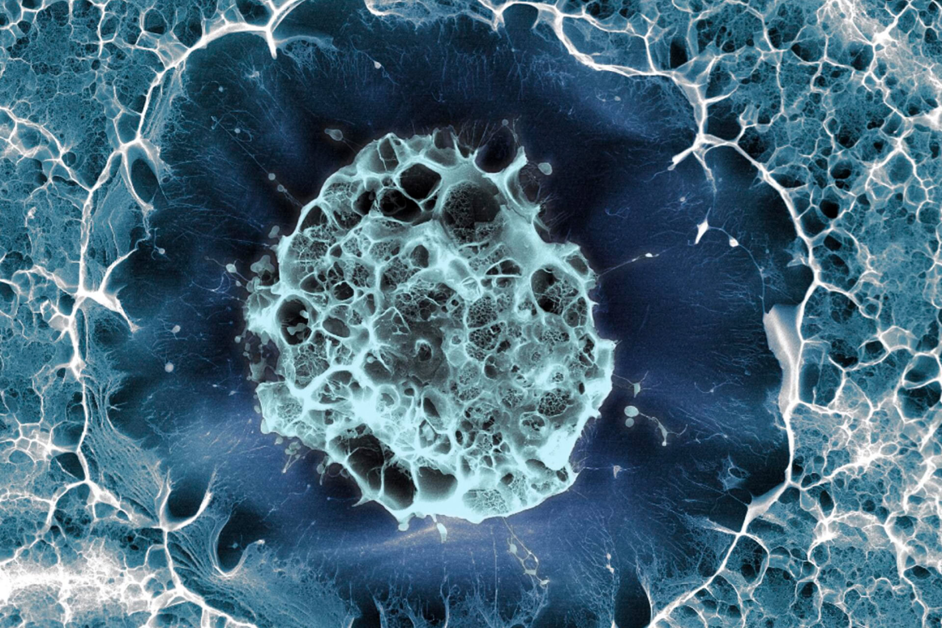
Details of how to develop three-dimensional organoid models of the cervix have been published.
An organoid is a small three-dimensional structure that is artificially developed to model whole organs. Researchers at the Julius Maximilians University of Würzburg, Germany aimed to mimic the complex interactions that take place in the cervix and replicate the conditions for tumorigenesis.
'Until recently, science has lacked a system that well reflects the cellular, physiological and functional properties of the different cell types in the cervix,' said Dr Cindrilla Chumduri, chair of microbiology at the University of Würzburg and the study lead.
It is widely accepted that the cervix is made up of two types of epithelial cell. Squamous epithelial cells are found in the outer part of the cervix and columnar epithelial cells are found in the inner part of the cervix, but Dr Chumduri and her team recently discovered that these cells are derived from two distinct stem cell populations, not one.
The lack of an in vitro system to model the cellular, physiological and functional properties of the two differing epithelial cells may have stalled previous efforts to effectively study cervix biology thus far. However, using adult epithelial stem cells as starting material, and a combination of growth factors and cultivation methods, the researchers were able to replicate the physical structure and functional properties of both cells. The scientists have published the procedure in Nature Protocols.
Using the two organoid models, they demonstrated that disrupting Wnt signalling alters the homeostasis of the cervix, causing one type of epithelium cell to replace the other. This event, known as metaplasia, can result in different types of cervical cancers depending on which cell population is displaced. These results were previously published in Nature Cell Biology (see BioNews 1080).
The scientists also genetically manipulated the stem cells by adding human papillomavirus oncogenes in order to model cervical cancer. As it is now believed that other factors increase the risk of cervical cancer, such as co-infection with other sexually transmitted diseases, including the bacterial disease Chlamydia trachomatis. These new organoids allow the scientists to study the effects of viral infection on the squamous epithelial cells of the cervix with the addition of co-infections with other pathogens, such as Chlamydia trachomatis.
'[These] organoids are ideal, almost physiological 3D epithelial tissues for studying and modelling cervical biology, host-pathogen interactions and disease development,' added Dr Chumduri. 'Organoids also make it possible to study the reaction of the cervical epithelium to hormonal changes and their effects on stem cell regeneration, mucus production and innate defence against pathogens. Their long-term cultivability offers a unique opportunity to take a closer look at chronic or repeated infections and their impact on host cells'.
The authors believe that gaining a better understanding of the biology of the cervix will improve our understanding of how bacterial and viral infections lead to cervical cancer, with applications in personalised medicine, genome editing, drug screening and disease modelling.
Sources and References
Related Articles
Bioprinted organoids to help predict cancer treatment response
A new method for the creation of tumour organoids has been developed, which allows them to be used with advanced imaging to study cancer in greater detail...

Podcast Review: Numberphile – The orchid room and cancer with Hannah Fry
The comfort provided by numbers, particularly for somebody with a knack for them, makes a lot of sense. However, as Professor Hannah Fry reveals in this episode of 'Numberphile', it is far more complicated than that...
Making bigger 'mini-organs' for research
Another breakthrough has been made towards the development of artificially-engineered tissue that mimics real organs that can be used for research...
Organoids give insight into the development of cervical cancer
Molecular changes that give rise to cervical cancer have been identified using novel 'organoid' models...
Leave a Reply
You must be logged in to post a comment.