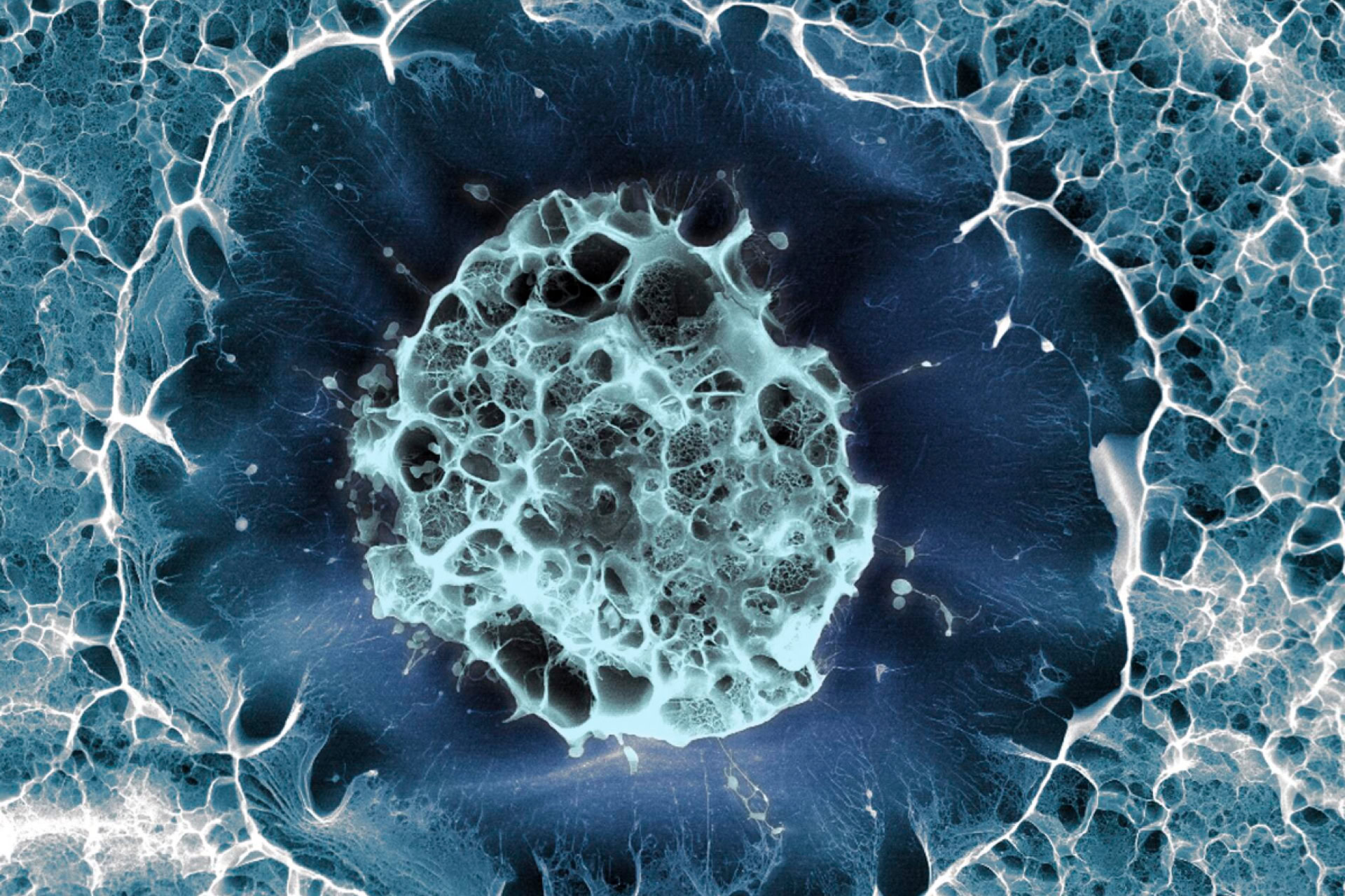
A new method for the creation of tumour organoids has been developed, which allows them to be used with advanced imaging to study cancer in greater detail.
Three-dimensional mini-tumours known as organoids can be created from patient's cells, or from cell lines, and exposed to therapeutic interventions to identify drug candidates. The effectiveness of those drugs can also be tested on the cells, thus helping choose the best therapy to treat patients. Yet, organoid methods cannot yet capture the diversity of cells that exist within the organoids. Some cells might respond well to the treatment while others do not, and current methods cannot discern these differences. Such diversity exists between cells within real tumours and can contribute to why treatments fail in patients.
Senior author of the study Dr Alice Soragni from the David Geffen School of Medicine at the University of California Los Angeles, said: 'Tumour organoids have become fundamental tools to investigate tumour biology and highlight drug sensitivities of individual patients… However, we still need better ways to anticipate if resistance could be arising in a small population of cells, which we may not detect using conventional screening approaches.'
In the new study published in Nature Communications, researchers attempted to address this limitation of organoids. They used a technique called bioprinting to precisely print the cells into uniform, thin layers of extracellular support proteins, forming three-dimensional tumours.
They then incorporated a method called high-speed live cell interferometry (HSLCI), which measures changes in the live cells' biomass and distribution in real time without being invasive or destructive to the organoid's development.
Organoid biomass is an important measure of its fitness, as it reflects the synthetic and degradative processes occurring within the cells. Using the HSLCI's measurements on the organoids, the researchers then used machine learning to analyse and classify the organoids.
The researchers confirmed they could accurately measure the growth patterns of the bioprinted tumour cells over time. They were also able to classify the organoids based on their response to different concentrations of a variety of drugs and to identify the response to the drugs within organoid samples, finding clusters of cells which respond less to the drug compared to the rest of the sample.
While this novel bioprinted organoid system combined with HSLCI and machine learning is currently in its conceptual stage, it does establish itself as a method that could be used in the future to model the response of tumours to treatments even more precisely.
'By using this method, we are able to accurately measure the masses of thousands of organoids simultaneously,' said Dr Michael Teitell, director of the UCLA Jonsson Comprehensive Cancer Centre and co-senior author of the study. 'This information helps identify which organoids are sensitive or resistant to specific therapies, which can be used to quickly select the most effective treatment options for patients.'


Leave a Reply
You must be logged in to post a comment.