

Human organoids transplanted into mouse brains can grow and form connections with the surrounding nervous system.
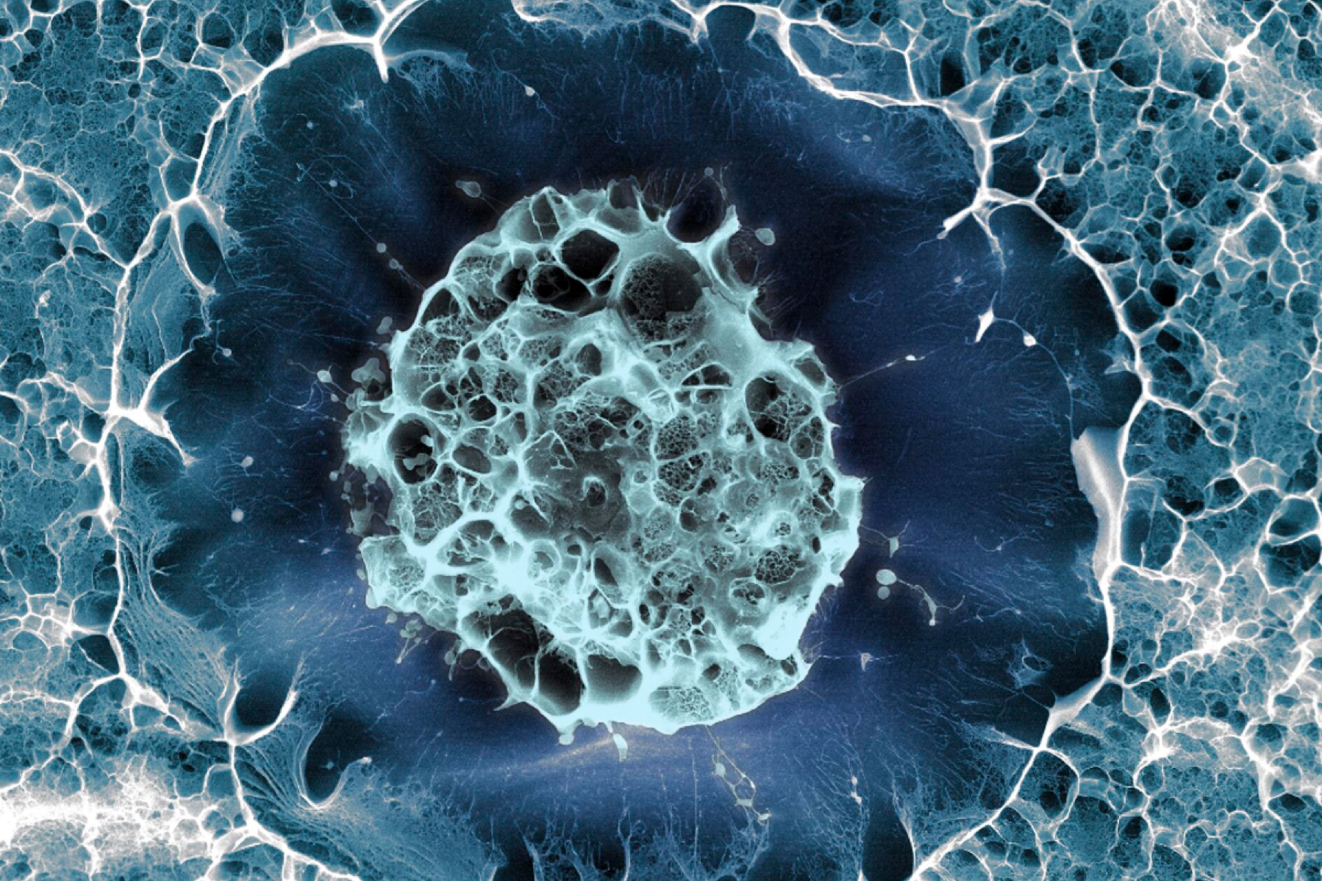
Human brain organoids are 3D structures grown in the laboratory which contain living, interconnected human neurons. They are made by reprogramming human skin cells into stem cells, which can develop into various neuronal cell types. Although organoids have been used in research for over a decade, they remain an in vitro model of the human brain. To overcome some of the limitations of this approach, a team from Stanford University, California, implanted human organoids into the brains of bioengineered mice that lack most of their cerebral cortex. This model allowed the researchers to investigate aspects of human brain development.
Professor Sergiu Pasca, senior author of this study and professor of psychiatry and behavioural sciences at Stanford University, said: 'These animal models offer a unique opportunity to study how disease-associated alterations in human brain circuitry manifest in an intact nervous system.' He added that, with existing organoid models: 'We couldn't study complex human behaviour in a dish.'
Previous studies have already implanted human brain organoids into living mice. However, this work, which was published in Nature, used bioengineered mice designed to accommodate human brain tissue. The researchers developed mice that were genetically engineered to lack most of the cerebral cortex and hippocampus. They implanted human cortical organoids derived from healthy donor cells, each containing around 100,000 cells, into the brains of two-day-old mice. Three months later, more than 90 percent by volume of the measured cortical tissue in the mice's brains was human-derived, with human neurons also extending through the mouse brain and as far as the spinal cord.
The team then explored how this model could be used to study human brain disease. The human tissue developed into specialised neurons, including rare von Economo neurons. These are particularly vulnerable in frontotemporal dementia and have never been grown in laboratory culture before.
The researchers also investigated neurological movement disorders. After exposure to low oxygen levels, similar to those that can occur around birth and are associated with cerebral palsy, only mice with human brain tissue developed motor coordination problems resembling those seen in people with the condition.
The research has also raised questions about the ethics of combining human brain tissue with animals. Professor Benjamin Hurlbut, a biosciences ethicist at Arizona State University who was not involved in the study, said the research is 'experimenting with concepts that are socially and morally meaningful, like the demarcation between human and nonhuman.' Professor Madeline Lancaster, a neurobiologist at the University of Cambridge who was also not involved, said: 'The goal here is clearly not to make a mouse that's super intelligent…[the goal is] so you can start using it to understand human neurobiology and human neurological diseases.'
Professor Pasca has stated that: 'Throughout several years of experimentation, we have received input from ethicists, neurobiologists with expertise in primate and human cortical biology, patient advocates, philosophers and legal scholars.' The team plans to use this model to investigate how human brain tissue responds to oxygen deprivation in cerebral palsy, as well as to study other conditions, such as schizophrenia and autism.
Sources and References
-
Stanford Medicine team creates advanced model for studying brain development, disorders
-
Developmental xenocortication using human-derived organoids in mice
-
Human brain cells transplanted into mice in 'most extensive' integration ever
-
Human neurons flourish in mouse brains, offering a new view of neurodevelopmental disorders
-
Scientists create mice with part-human brains
-
Part-human part-mouse brain developed in science breakthrough
Related Articles

Salivary gland organoids developed from human pluripotent stem cells
Human salivary gland organoids derived from pluripotent stem cells have been transplanted into mice....

Human brain develops from two distinct groups of cells
The origins of the human brain can be traced back to two distinct groups of cells in the developing embryo...
Human brain organoids kept for more than five years in a dish
Human brain organoids have been kept in culture for more than five years, extending the period over which these models of human brain development have previously been maintained...
Brain organoids could guide treatment of Alzheimer's disease
Brain organoids, and the tiny particles they secrete, may help to stage early Alzheimer's disease and evaluate drug response...
Brain organoids help identify Alzheimer's drug target
A potential new gene target for Alzheimer's disease treatment has been identified using brain organoids...
Whole-brain organoid mimics brain of 40-day-old fetus
Organoids comprising brain and blood vessel cells have been generated from human pluripotent stem cells, yielding a composition of different cell types similar to that of a 40-day-old fetus...
Mice, pigs and humans: chimaera research at the ISSCR conference
Novel types of chimaeric pig embryos and chimaeric mice, containing both human and animal cells, have been reported at an international conference...
Chimeric brain organoid created using stem cells from five people
Stem cell lines from multiple human donors have been combined to create 3D brain organoids for the first time...