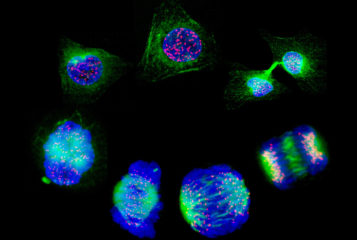
Image by NIMR/Francis Crick Institute via the Wellcome Collection. Depicts cultured cells stained to show the nucleus in blue, the tubulin in green and the mitochondria in red.
382 articles
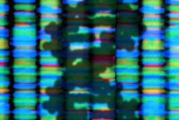
Image by Peter Artymiuk via the Wellcome Collection. Depicts the shadow of a DNA double helix, on a background that shows the fluorescent banding of the output from a DNA sequencing machine.
Image by Peter Artymiuk via the Wellcome Collection. Depicts the shadow of a DNA double helix, on a background that shows the fluorescent banding of the output from a DNA sequencing machine.
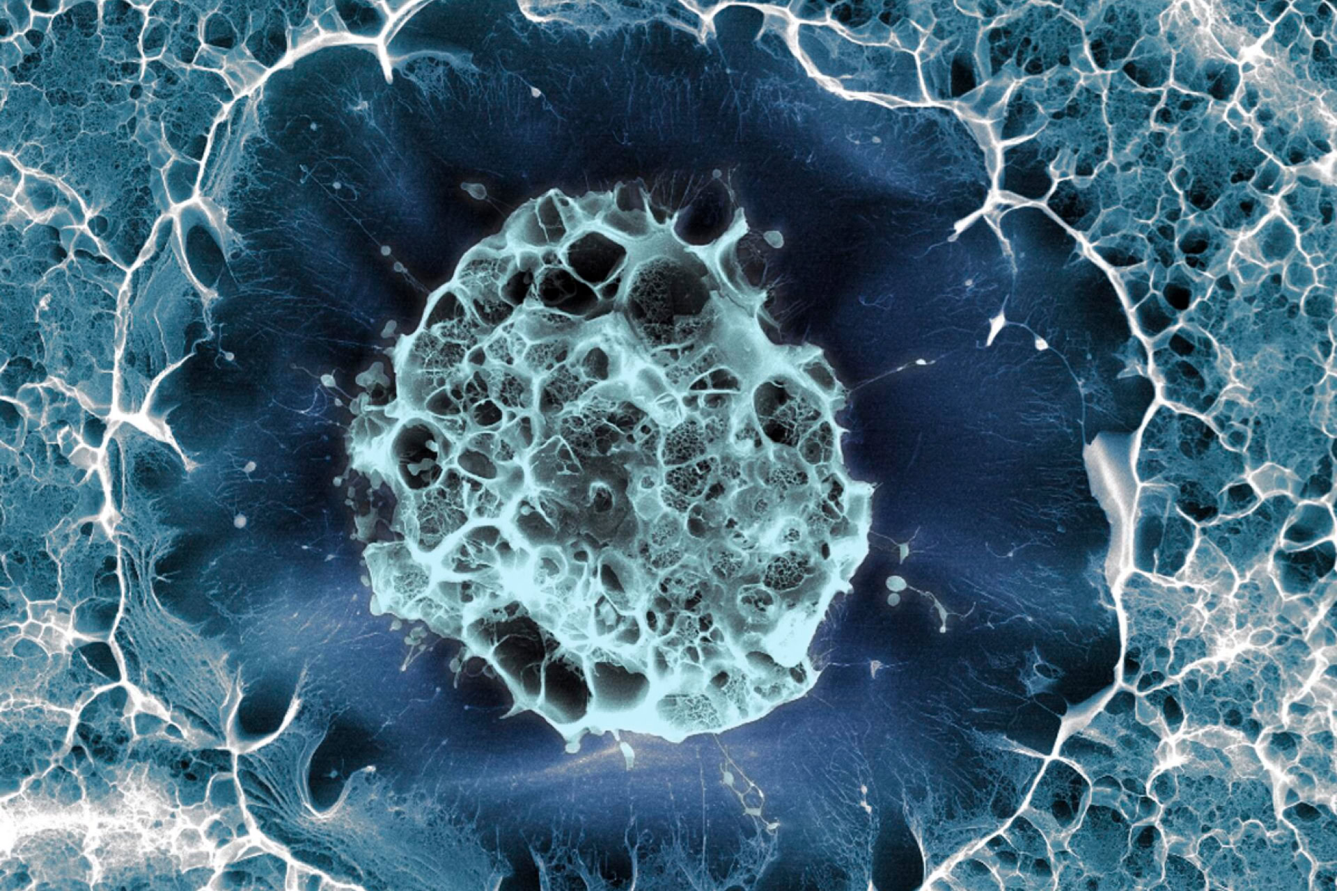
Image by Sílvia Ferreira, Cristina Lopo and Eileen Gentleman via the Wellcome Collection. Depicts a single human stem cell embedded within a porous hydrogel matrix (false colour).
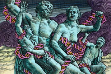
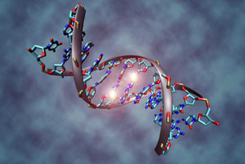
Image by Christoph Bock/Max Planck Institute for Informatics via Wikimedia Commons. Depicts a DNA molecule that is methylated on both strands on the centre cytosine.
Image by Peter Artymiuk via the Wellcome Collection. Depicts the shadow of a DNA double helix, on a background that shows the fluorescent banding of the output from a DNA sequencing machine.
Image by Peter Artymiuk via the Wellcome Collection. Depicts the shadow of a DNA double helix, on a background that shows the fluorescent banding of the output from a DNA sequencing machine.
Subscribe to BioNews and other PET updates for free
BioNews, published by the Progress Educational Trust (PET), provides news and comment on genetics, assisted conception, embryo/stem cell research and related areas.