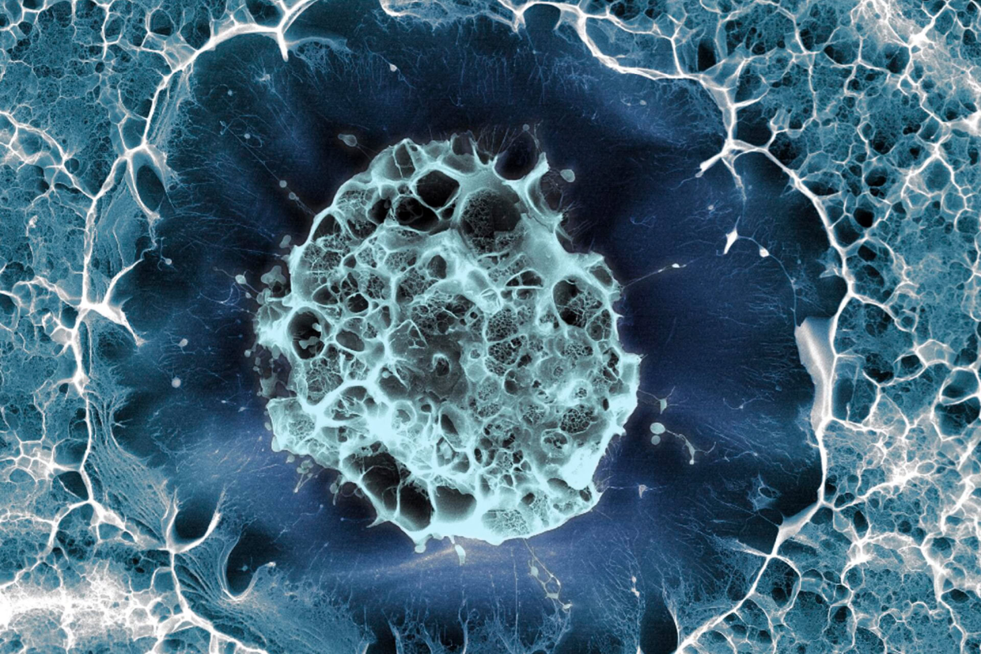
Brain organoids made from human stem cells can integrate successfully with the visual system in rats, offering hope for repairing brain damage.
Brain organoids are 3D structures made from stem cells that work like a model of the brain. Sometimes called a 'brain in a dish', they have been used to study brain development, disease, injury, and to test potential treatments. However, this study is the first to demonstrate that human brain organoid grafts can integrate with living injured adult mammalian brains.
Dr Han-Chiao Isaac Chen who led the research at the University of Pennsylvania's Department of Neurosurgery said, 'Brain organoids have architecture; they have a structure that resembles the brain. We were able to look at individual neurons within this structure to gain a deeper understanding of the integration of transplanted organoids.'
In the study, published in Cell Stem Cell, the group grew human pluripotent stem cell lines in a laboratory dish to create organoids. Next, these were grafted into the brains of rats with damaged visual cortexes. Immunosuppression medication was used to prevent the grafts from being rejected.
The study reported a graft survival rate of 82 percent and noted that astrocytes (a critical and highly specialised neural cell type) were more prevalent in the host brain near the organoid graft. The density of the new neural projections also increased in important brain regions such as the hippocampus and the motor complex; the targets of the projections were primarily part of the visual system.
The researchers traced the connections between the grafted brain organoids and the host brain cells of the rats by injecting the eye with a fluorescently tagged virus. These modified viruses can cross synapses (junctions between nerve cells) and jump from neuron to neuron allowing the researchers to track them visually and see pathways of connections.
They then used electrode probes to measure the electrical activity of the individual neurons in the brain organoid while exposing the rats to flashing lights and alternating white and black bars. They found that several neurons in the organoid responded to the stimulus, showing that the organoid neurons were integrating with the visual system and adopting the functions of the visual cortex.
Most previous studies attempting to transplant neurons into existing brains have used injections of many individual cells, rather than cells already in an ordered and complex structure. Although the study does not model any specific disorder or brain injury, it shows the potential for organoid integration with the host brain. The authors note that this will not be the optimal integration – which may require significant maturation of the graft neurons – meaning that better results may be achievable with more research. They also want to repeat the research by looking at other brain areas, such as the motor cortex.
'We hope this study moves us in the direction of restoring function using these organoids and eventually leads to, in the long term, transplanting organoids into patients with brain injuries,' said Dr Chen.
Sources and References
-
Human neurons implanted into a rat's brain respond to flashing lights
-
Structural and functional integration of human forebrain organoids with the injured adult rat visual system
-
Transplanted human brain cells respond to visual stimuli in rat brains
-
Human stem cell-derived tissue transplanted into rats show potential for repairing damaged brains
-
Human 'mini brains' merge with injured rat brain tissue and respond to light


Leave a Reply
You must be logged in to post a comment.