Brain organoids developed from fetal tissue offer a new way to model brain diseases and aid in drug testing.
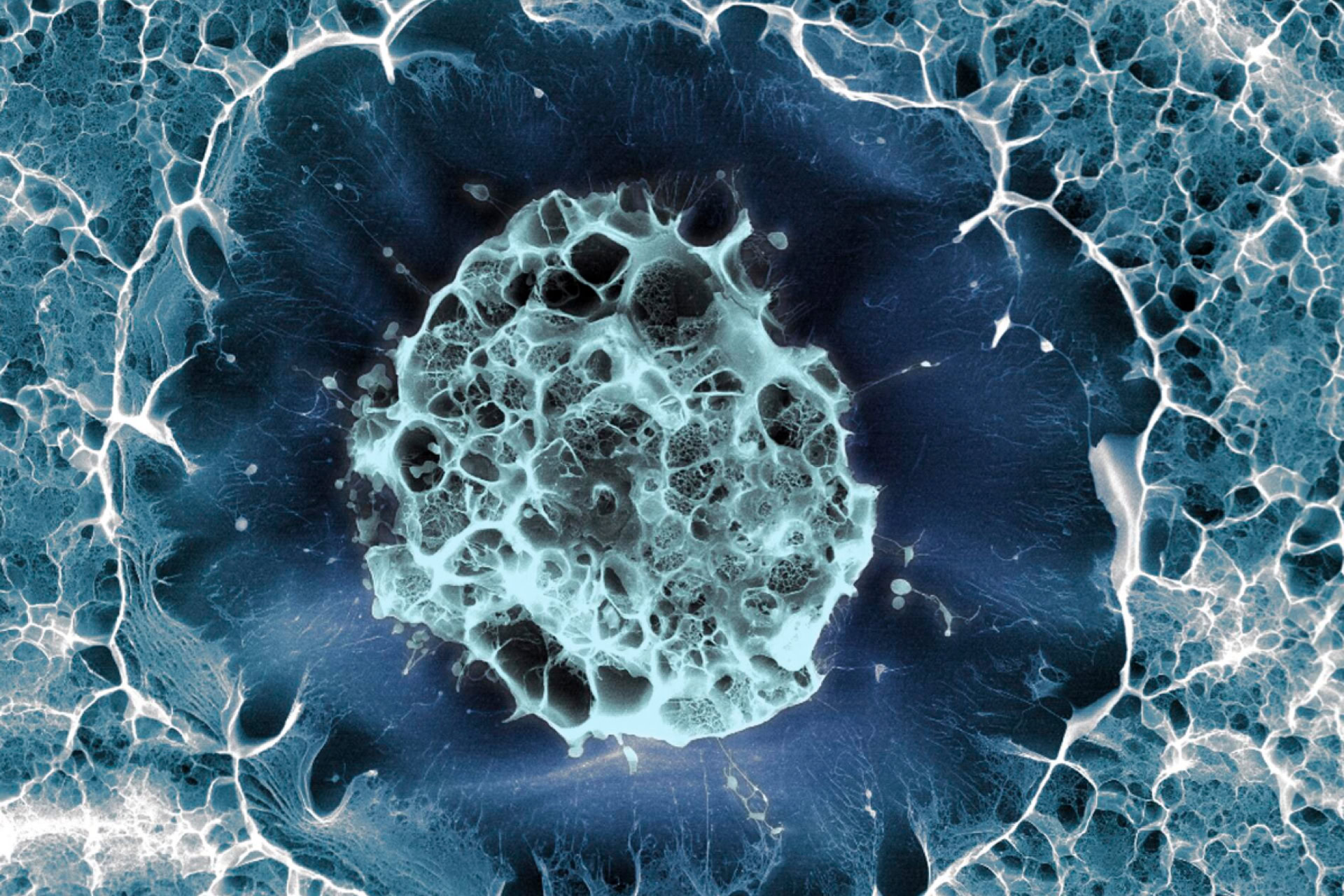
Organoids are 3D structures which closely mimic actual tissue, enabling the study of human organs in health and disease. Brain organoids have previously been made from human stem cells directed by a cocktail of molecules in order to mimic the development of the brain (see BioNews 1177). Authors in this study, published in Cell, have shown that small pieces of fetal brain tissue could self-organise into brain organoids, which may be used to model brain diseases, such as developmental conditions and brain cancers, and also aid in drug testing.
'Brain organoids from fetal tissue are an invaluable new tool to study human brain development. We can now more easily study how the developing brain expands, and look at the role of different cell types and their environment,' said Dr Benedetta Artegiani, research group leader at the Princess Máxima Centre in the Netherlands, who co-led the research.
'Our new, tissue-derived brain model allows us to gain a better understanding of how the developing brain regulates the identity of cells. It could also help understand how mistakes in that process can lead to neurodevelopmental diseases such as microcephaly, as well as other diseases that can stem from derailed development, including childhood brain cancer.'
The researchers collected tissue from three different regions, including the forebrain, cortex and spinal cord, of 15-week-old fetal brains and cut them into pieces before placing into dishes with molecules used to trigger growth.
The fetal brain organoids, around the size of a grain of rice, successfully recapitulated the original tissue they were derived from and produced a kind of 'scaffolding' around cells called the extracellular matrix. By changing the molecules that surrounded organoids, they could be matured showing increased expression of markers representative of a more developed brain.
The genome editing approach CRISPR/Cas9 was then used to create a model of brain cancer, showcasing the multitude of ways these organoids can be useful for the scientific community in the future.
The tissue was obtained from aborted fetuses. The donors whose pregnancies had been terminated had to provide informed consent before any fetal tissue could be used in research, and these donors were not compensated. The researchers worked with bioethicists to provide assurance that the fetal organoids could not feel pain or become conscious, outcomes which were deemed unlikely due to the lack of sensory inputs or interactions between mature brain regions.
Professor Alysson Muotri – a human brain organogenesis specialist from the University of California, San Diego, who was not involved in the work – told Science 'It serves as a validation that [stem-cell–derived] organoids are really mimicking the fetal brain,'.
Sources and References
-
New organoids could revolutionise brain research
-
Human fetal brain self-organizes into long-term expanding organoids
-
First brain organoids grown from fetal tissue offer window on development
-
Brain organoids from fetal tissue take CRISPR changes, enable tumour modeling
-
Long-term expansion and self-organisation of human fetal brain organoids


Leave a Reply
You must be logged in to post a comment.