A group of Australian scientists has used a new genetic analysis technique to assess IVF embryos, to identify those most likely to develop in the womb. The findings were published in the journal Human Reproduction last week.
Around one per cent of all births in the UK are from IVF treatment and the latest figures show that of 32,600 women who underwent treatment, only 11,000 resulted in births. One in four IVF pregnancies is multiple, compared with one in 80 for a natural conception.
The process of IVF involves extracting many eggs for fertilisation before choosing one, two or sometimes even three embryos for transfer into the uterus. This can lead to multiple pregnancies, which carry extra health risks for mothers and babies. Embryo selection is currently based on observations of morphology (shape and appearance) to predict their potential viability. The problem could be overcome by finding an objective, measurable means of testing embryo viability, rather than a subjective one such as morphology, to definitively pick a single, viable embryo.
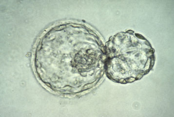
The study, carried out by scientists at the Monash Immunology and Stem Cell Laboratories, Monash University, Australia, and the Centre for Human Reproduction, Genesis Athens Hospital, Athens, Greece, recruited 48 women undergoing IVF treatment. Once their fertilised eggs reached the 'blastocyst' stage, an early stage of development around day five, between eight and 20 cells from the surface layer were removed. These samples were then genetically analysed using microarray techniques, which measures gene activity in the cells.
Of the embryos selected as being viable, one or more were transferred into the 48 women, 25 of whom became pregnant, with 37 babies being born. The scientists took DNA samples from the babies, and used DNA fingerprinting to match which blastocyst grew into which baby. This enabled them to compare the activity levels of key genes, to identify which had been active in the viable, compared to the non-viable early embryos. These genes identified were involved in key processes such as cell adhesion, cell communication, cellular metabolic processes and response to stimuli.
The research yielded some important findings; not only that such analysis can be used to distinguish between viable and non-viable embryos from IVF by looking at gene activity patterns, but also that up to 20 surface layer cells can be extracted from a blastocyst without adversely affecting its viability.
Dr Gayle Jones, the lead co-author and senior research scientist at Monash University, said: 'the ability to select the single most viable embryo from within a cohort available for transfer will revolutionise the practice of IVF, not only improving pregnancy rates but eliminating multiple pregnancies and attendant complications'. Her aim is to eventually identify a subset of five to ten genes that 'could be put into a very rapid same day test for use in IVF clinics'.


Leave a Reply
You must be logged in to post a comment.