New research suggests COVID-19 can infect and multiply within intestinal cells.
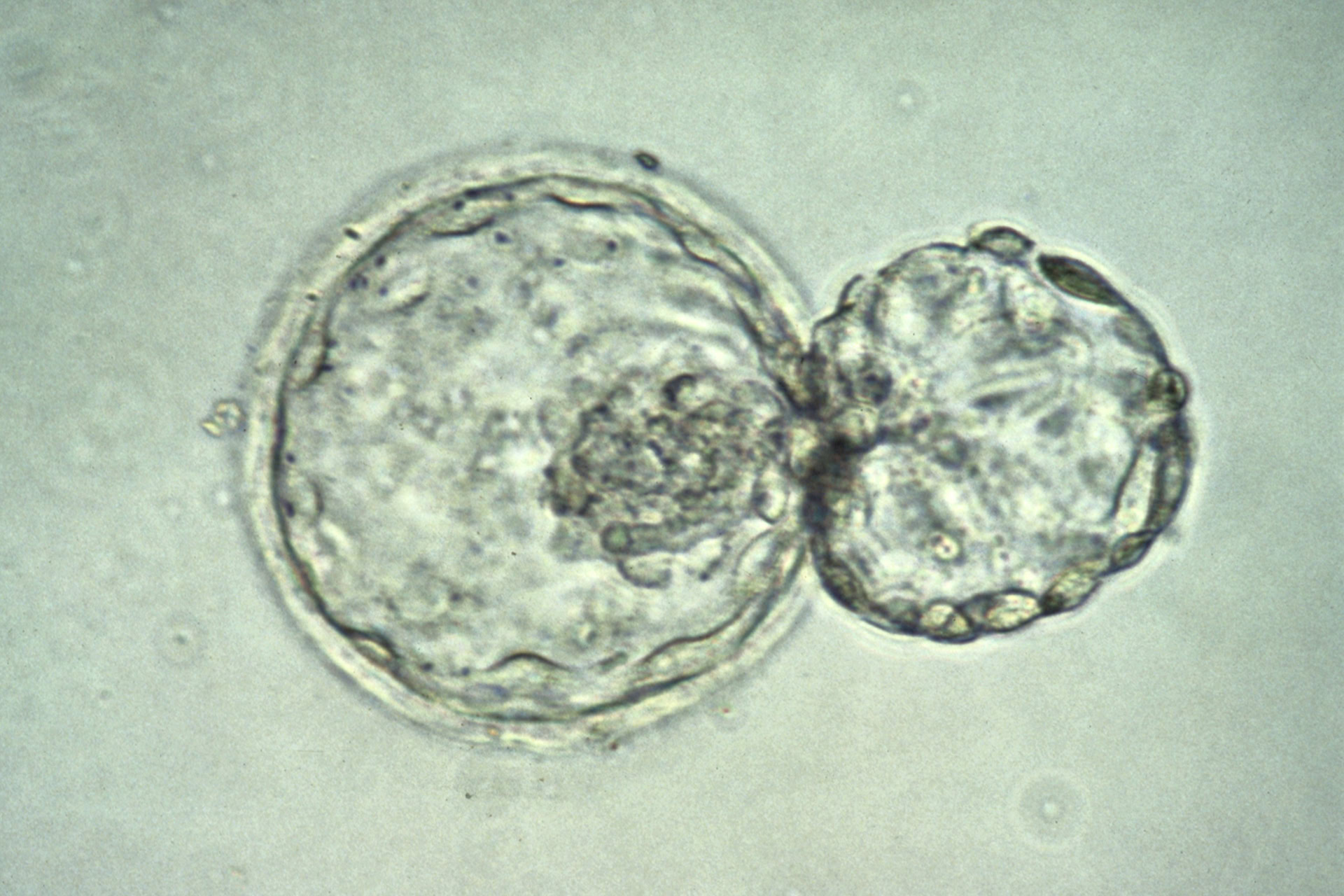
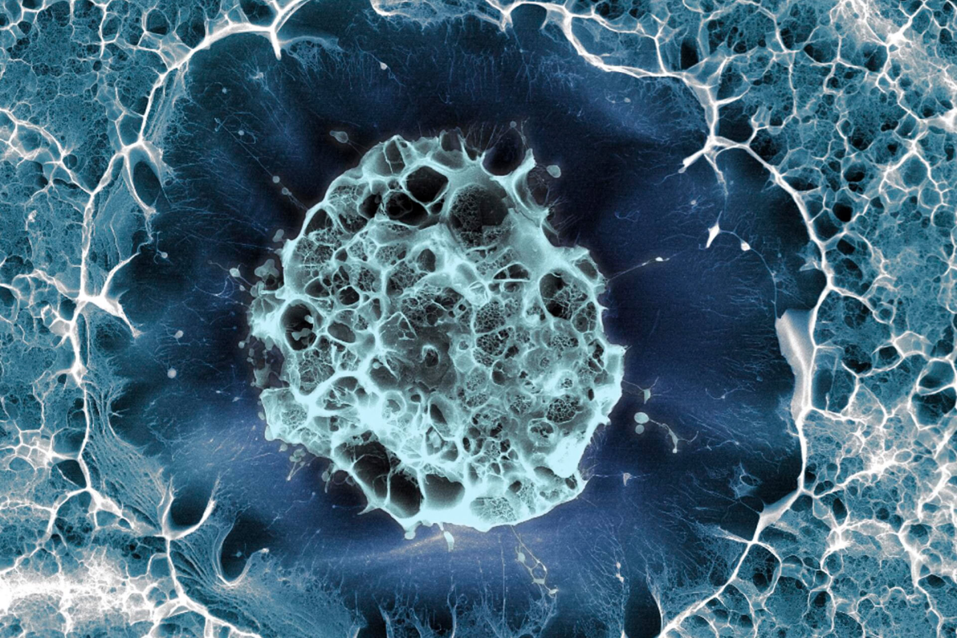
Researchers, based at the Hubrecht Institute, Erasmus MC University Medical Centre and Maastricht University in the Netherlands, used three-dimensional models of the intestine grown in the lab. Their findings may explain how the virus is often detected in stool samples, and why a third of COVID-19 patients experience gastrointestinal symptoms.
'These organoids contain the cells of the human intestinal lining', Professor Hans Clevers, a lead scientist on the study explained, which 'makes them a compelling model to investigate infection by SARS-CoV-2'.
Like respiratory organs, cells on the inside of the intestine have ACE2, the receptor via which COVID-19 enters cells. However, until now, it was unknown whether this meant intestinal cells could be infected and produce viral particles.
The scientists exposed the 3D-structures to the virus, and measured its spread using microscopy techniques. Their findings, published in Science, show that the virus-infected cells are of the enterocyte lineage, which makes up absorptive cells lining the inner surface of the intestine.
The team then measured the activation of genes within these cells in response to the virus using RNA sequencing, finding that genes 'known to fight viral infections' are triggered. 'Future research will focus more closely on these genes, and how they might be used to develop new treatments', the researchers said.
However, their results do not show whether intestinal COVID-19 plays a role in transmission of the disease. 'Our findings indicate that we should look into this possibility more closely', noted Professor Bart Haagmans, another lead scientist on the study. In the meantime, their research efforts continue by comparing lung and intestinal organoids infected with the virus.


Leave a Reply
You must be logged in to post a comment.