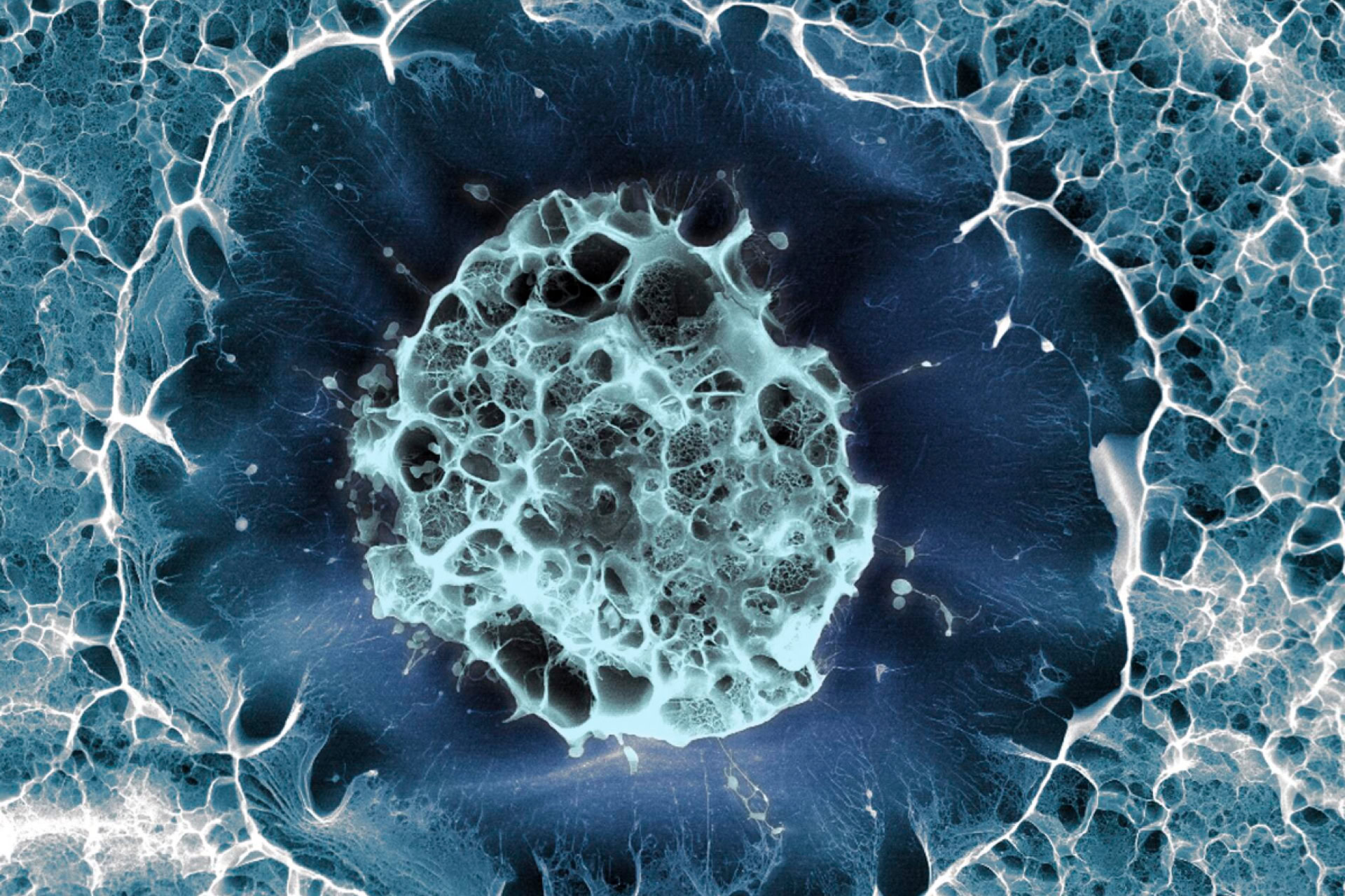
Two teams of researchers have successfully grown 3D lung models to study the progression of COVID-19.
The models allow researchers to study the infection of lung cells with SARS-CoV-2, the virus which causes COVID-19, in real time. The lung models, called organoids, could also be used to screen new therapies for treating COVID-19.
'We still know surprisingly little about how SARS-CoV-2 infects the lungs and causes disease,' explained joint senior author of one of the studies, Dr Joo-Hyeon Lee of the Wellcome-MRC Cambridge Stem Cell Institute, University of Cambridge.
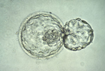
'Our approach has allowed us to grow 3D models of key lung tissue – in a sense, "mini-lungs" – in the lab and study what happens when they become infected,' she added.
Two studies were published separately in the journal Cell Stem Cell. One was conducted largely in the UK, at the University of Cambridge, in conjunction with researchers from the Korea Advanced Institute of Science and Technology (KAIST) in Seoul, South Korea. The second study was carried out by researchers at Duke University in Durham, North Carolina, and the University of North Carolina at Chapel Hill. Both teams independently reached the same conclusions.
The studies build on a technique published earlier this year, which describes growing organoids with purely human cells (see BioNews 1058). Each group isolated a type of cell called AT2s, or alveolar type 2 cells, from healthy human lung structures called alveoli – small air sacs which exchange carbon dioxide with oxygen as we breathe. AT2s are a specialised form of stem cell capable of dividing and growing into different lung cell types, but are also susceptible to SARS-CoV-2 infection. The researchers grew these cells into 3D organoids and infected them with SARS-CoV-2.
Both teams found that shortly after infection, AT2 cells began producing molecules called interferons – a distress signal to the immune system and a warning to neighbouring cells. The researchers also discovered that lung stem cells could themselves activate the innate immune system, the first line of defence against infection, in under 48 hours. By 60 hours post-infection, both infected and non-infected cells had begun to die, causing tissue damage.
Each research group also compared changes in genes of the infected organoids with those seen in the lungs of COVID-19 patients, and found that they were remarkably similar – suggesting that the organoids are a reliable model for studying SARS-CoV-2 infection.
The findings have wide-reaching implications for COVID-19 research. For instance, by using AT2 cells from different donors, researchers can study the impact of factors such as age on COVID-19 progression.
'Based on our model we can tackle many unanswered key questions,' claimed joint senior author Dr Young Seok Ju, from KAIST. He added: 'Most importantly, it [the model] provides the opportunity to develop and screen potential therapeutic agents against SARS-CoV-2 infection.'
Senior author of the alternate study, Dr Purushothama Rao Tata at Duke University, believes the implications of the model go beyond COVID-19 research. He said: 'This is a versatile model system that allows us to study not only SARS-CoV-2, but any respiratory virus that targets these cells, including influenza.'
Sources and References
-
Lab-grown mini-lungs mimic the real thing – right down to COVID infection
-
Human lung stem cell-based alveolospheres provide insights into SARS-CoV-2 mediated interferon responses and pneumocyte dysfunction
-
'Mini-lungs' reveal early stages of SARS-CoV-2 infection
-
Three-dimensional human alveolar stem cell culture models reveal infection response to SARS-CoV-2
-
Scientific breakthrough as researchers use stem cells to grow lungs
-
'Mini-lungs' reveal early stages of COVID-19 infection
Related Articles
Organoid models of paediatric liver cancer developed
Model livers, which contain cells that are genetically identical to tumour cells, have been developed to study a rare paediatric liver cancer...
Researchers grow mini human lungs to imitate COVID-19 infection
The first human 'lung-in-a-dish' model, known as a lung organoid, representing all lung cell types and allowing replication of SARS-CoV-2 infection has been produced...
Airway model developed to study viral lung infections
Stem cell models may help investigate COVID-19 and other lung diseases...
Organoids of human lung and brain respond differently to SARS-CoV-2
The body's response to infection by SARS-CoV-2 (the virus that causes COVID-19) varies across different cell types, according to a new study...
Making bigger 'mini-organs' for research
Another breakthrough has been made towards the development of artificially-engineered tissue that mimics real organs that can be used for research...
Studying SARS-CoV-2 using human lung organoids
Scientists from Stanford University, California, USA have developed a new model system to better study the lung damage that occurs during COVID-19...
Mini organs show how coronavirus attacks the body
Mini organs are being developed from stem cells to study how the SARS-Co-V2 affects the body...
Organoid models suggest COVID-19 virus infects human intestinal cells
New research suggests COVID-19 can infect and multiply within intestinal cells...
Leave a Reply
You must be logged in to post a comment.