PET PET
  • My Account
  • Subscribe
Become a Friend Donate
  • About Us
    • People
    • Press Office
    • Our History
  • Get Involved
    • Become a Friend of PET
    • Volunteer
    • Campaigns
    • Writing Scheme
    • Partnership and Sponsorship
    • Advertise with Us
  • Donate
    • Become a Friend of PET
  • BioNews
    • News
    • Comment
    • Reviews
    • Elsewhere
    • Topics
    • Glossary
    • Newsletters
  • Events
    • Upcoming Events
    • Previous Events
  • Engagement
    • Policy and Projects
      • Resources
    • Education
  • Jobs & Opportunities
  • Contact Us
  • My Account
  • Subscribe
  • Privacy Statement
  • Advertising Policy
  • Thanks and Acknowledgements
PETBioNewsCommentSeeing in 3D

BioNews

Seeing in 3D

Published 29 July 2013 posted in Comment and appears in BioNews 715

Author

Dr Dusko Ilic

Image by K Hardy via the Wellcome Collection. Depicts a human embryo at the blastocyst stage (about six days after fertilisation) 'hatching' out of the zona pellucida.
CC BY 4.0
Image by K Hardy via the Wellcome Collection. Depicts a human embryo at the blastocyst stage (about six days after fertilisation) 'hatching' out of the zona pellucida.

We've all heard the stories about Archimedes taking a bath, Newton sitting under an apple tree - about moments when the secrets of nature suddenly revealed themselves to humankind. Well, stem cell science and regenerative medicine are nothing like that!...

We've all heard the stories about Archimedes taking a bath, Newton
sitting under an apple tree - about moments when the secrets of nature suddenly
revealed themselves to humankind. Well, stem cell science and regenerative
medicine are nothing like that!

Every success is built on years and years of painstaking research,
on numerous small but important steps, each essential for us to move forward. A
paper in Nature Biotechnology from Professor Robin Ali and groups at University
College London, Institute of Ophthalmology and Moorfields Eye Hospital, is one such
step.

It was not a spur of the moment development; it was built on Professor
Ali's previous work and on a new laboratory technique of three-dimensional (3D)
culture developed by Dr Yoshiki Sasai at the RIKEN Center for Developmental
Biology in Japan. Protocols involving pluripotent stem cells, both embryonic and induced, had already been developed, but no-one had
successfully used stem cells as a source of photoreceptor cells suitable for
retinal transplantation. This study did it for the first time.

Would results of this study return eyesight to the blind? Sadly not, so
why did this make such a big news splash? The study is an important milestone. Professor
Ali and his team realised a while ago that a traditional 2D cell culture system
is not capable of providing the right environment for differentiation of
pluripotent stem cells into photoreceptor precursors. There was something
missing.

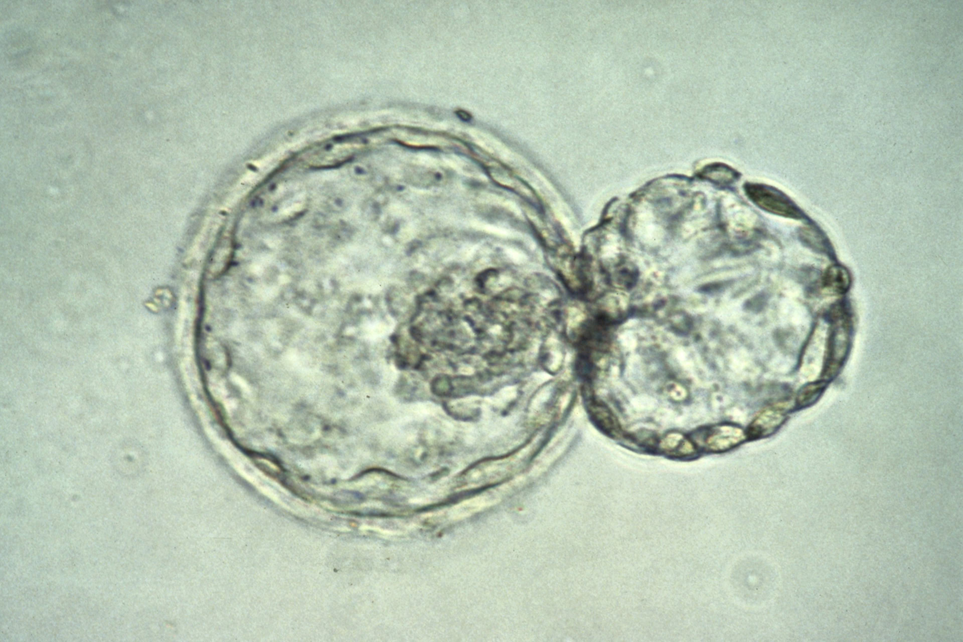
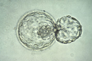
They hoped that they might circumvent the problem if they were able
to copy the microenvironment of embryonic tissue during development. Work
from Dr Sasai's lab suggested that introducing 3D cell culture was the best way
to go. And indeed it was. Retinal progenitor cells generated in the 3D system
were capable of differentiation into mature retinal cell types. Moreover, these
cells were able to integrate with the mouse retina. The transplanted cells,
made distinguishable through expression of green fluorescent protein, were
detected during follow-up six weeks after transplantation.

The study has broader implications than the step toward restoration
of vision through transplantation of photoreceptors. It points out how
important it is to mimic closely the natural microenvironment.

It has been known for many years that culturing embryos under
physiological oxygen levels yields much better results than culturing them at
normal atmospheric levels. For dermatologists building a 3D epidermis, only
exposure of the skin cells to atmospheric air will result in the proper
stratification of epidermis. A couple of weeks ago, scientists in Japan grew
functional liver buds
in vitro (see BioNews 712). They mixed together hepatocyte progenitors
derived from human pluripotent stem cells, human umbilical vein endothelial
cells and human mesenchymal cells - three cell types normally present in the liver
— and cultured them together. The results were astonishing; the researchers were
able to rescue animal models of liver failure by transplanting such off-the-shelf
liver buds. Dr Hideki Taniguchi, who led the team, shared the same vision as Robin
Ali: mimic the natural microenvironment as closely as possible.

But will these moves to mimic
nature usher in a new era of wizardry, where we will be cooking up witches'
brews recipes of dead toads and bats, cell of mesenchyme and tissue of embryo, to
grow a new heart or brain in a dish? No. In order to manipulate a system and
get what we want when we want, we have to learn the molecular mechanisms and
understand the laws of nature.

Just remember, Judah Folkman, the founder of the field of angiogenesis, was the first to note that growth of all malignant tumours is
angiogenesis-dependent. However, the discoveries that originated the concept
and developed the field were not enough to develop the first potent therapeutic
antiangiogenic agent. It is rather Napoleone Ferrara, who identified the human
vascular endothelial growth factor (VEGF) gene and described its proangiogenic
characteristics, who is credited for leading the way to the first
antiangiogenic therapy, bevacizumab (Avastin).

Who will win the race to provide off-shelf
organs or organoids in the field of regenerative medicine? History suggests it won't
be the groups whose work has got us this far, nor those who'll continue the
fine-tuning. So although the work from Professor Ali's group is undeniably a milestone,
there are many more steps to come before this strategy could be used as a cell-based
therapy for particular types of blindness.

Related Articles

Image by K Hardy via the Wellcome Collection. Depicts a human embryo at the blastocyst stage (about six days after fertilisation) 'hatching' out of the zona pellucida.
CC BY 4.0
Image by K Hardy via the Wellcome Collection. Depicts a human embryo at the blastocyst stage (about six days after fertilisation) 'hatching' out of the zona pellucida.
News
30 March 2016 • 2 minutes read

3D bioprinter builds artificial bone, muscle and cartilage

by Kulraj Singh Bhangra

Researchers in the USA have designed a 3D printer that can build living cells around a biodegradable structure to construct various artificial tissues...

Image by K Hardy via the Wellcome Collection. Depicts a human embryo at the blastocyst stage (about six days after fertilisation) 'hatching' out of the zona pellucida.
CC BY 4.0
Image by K Hardy via the Wellcome Collection. Depicts a human embryo at the blastocyst stage (about six days after fertilisation) 'hatching' out of the zona pellucida.
News
21 August 2015 • 2 minutes read

Most complete 'human brain' grown in lab

by Dr Julia Hill

For the first time, an almost fully formed human brain has been grown in the lab, according to scientists from Ohio State University...

Image by K Hardy via the Wellcome Collection. Depicts a human embryo at the blastocyst stage (about six days after fertilisation) 'hatching' out of the zona pellucida.
CC BY 4.0
Image by K Hardy via the Wellcome Collection. Depicts a human embryo at the blastocyst stage (about six days after fertilisation) 'hatching' out of the zona pellucida.
News
6 March 2013 • 2 minutes read

Eye precursor and tiny liver grown from stem cells

by Dr Daniel Grimes

Human embryonic stem cells have, for the first time, been used to grow a crucial part of the eye, a paper in Cell Stem Cell reports. It is hoped that in the future transplantation of such tissue could help visually impaired people recover their sight...

Image by Peter Artymiuk via the Wellcome Collection. Depicts the shadow of a DNA double helix, on a background that shows the fluorescent banding of the output from a DNA sequencing machine.
CC BY 4.0
Image by Peter Artymiuk via the Wellcome Collection. Depicts the shadow of a DNA double helix, on a background that shows the fluorescent banding of the sequencing output from an automated DNA sequencing machine.
Comment
30 January 2013 • 3 minutes read

With an eye to the future - preliminary results of clinical trial in human embryonic stem cell-based therapy of macular degeneration

by Dr Dusko Ilic and 1 others

Last week, Advanced Cell Technology (ACT) of Massachusetts, USA, made two important announcements regarding human embryonic stem (hES) cell-based therapies for the potential treatment of Stargardt's dystrophy and age-related macular degeneration, two devastating degenerative disease leading to blindness....

Image by K Hardy via the Wellcome Collection. Depicts a human embryo at the blastocyst stage (about six days after fertilisation) 'hatching' out of the zona pellucida.
CC BY 4.0
Image by K Hardy via the Wellcome Collection. Depicts a human embryo at the blastocyst stage (about six days after fertilisation) 'hatching' out of the zona pellucida.
News
25 October 2012 • 2 minutes read

Mouse retina grown from stem cells

by Dr Rachael Panizzo

Embryonic stem cells have been used to generate a basic retina, the part of the eye that detects light and is needed for vision. The retinal tissue could be used to treat some forms of blindness, such as retinitis pigmentosa and age-related macular degeneration, and to investigate and screen potential new drugs for a range of eye diseases....

Image by Sílvia Ferreira, Cristina Lopo and Eileen Gentleman via the Wellcome Collection. Depicts a single human stem cell embedded within a porous hydrogel matrix (false colour).
CC BY 4.0
Image by Sílvia Ferreira, Cristina Lopo and Eileen Gentleman via the Wellcome Collection. Depicts a single human stem cell embedded within a porous hydrogel matrix (false-coloured cryogenic scanning electron micrograph).
News
9 June 2009 • 2 minutes read

Vision restored in mice using later stem cells

by Heidi Nicholl

In groundbreaking research, scientists have used a new approach to stem cell science to partially restore vision in mice genetically engineered to mimic human degenerative sight defects such as retinitis pigmentosa and macular degeneration. The team, a collaboration between University College London and the University of Michigan...

Leave a Reply Cancel reply

You must be logged in to post a comment.

« Old fertility science is still painfully relevant for today's women

Data-Label The UK's Leading Supplier Of Medical Labels & Asset Labels

RetiringDentist.co.uk The UK's Leading M&A Company.

Find out how you can advertise here
easyfundraising
amazon

This month in BioNews

  • Popular
  • Recent
8 August 2022 • 2 minutes read

FILM: 200 Years of Mendel – From Peas to Personalised Medicine

1 August 2022 • 4 minutes read

Women's Health Strategy plans reflect rising needs of same-sex female couples

25 July 2022 • 4 minutes read

Was the Women's Health Strategy worth the wait?

25 July 2022 • 4 minutes read

Why the UK should extend the 14-day rule to 28 days

25 July 2022 • 5 minutes read

200 Years of Mendel: From Peas to Personalised Medicine

15 August 2022 • 5 minutes read

Same-sex parent should not have been forced to adopt child

15 August 2022 • 2 minutes read

FILM: Editing the Human Genome – Where Are We Now? What Happens Next?

8 August 2022 • 4 minutes read

Citizenship and same-sex parents – about time, Sweden!

8 August 2022 • 2 minutes read

FILM: 200 Years of Mendel – From Peas to Personalised Medicine

1 August 2022 • 4 minutes read

Women's Health Strategy plans reflect rising needs of same-sex female couples

Subscribe to BioNews and other PET updates for free.

Subscribe
  • Twitter
  • Facebook
  • Instagram
  • LinkedIn
  • YouTube
  • RSS
Wellcome
Website redevelopment supported by Wellcome.

Website by Impact Media Impact Media

  • Privacy Statement
  • Advertising Policy
  • Thanks and Acknowledgements

© 1992 - 2022 Progress Educational Trust. All rights reserved.

Limited company registered in England and Wales no 07405980 • Registered charity no 1139856

Subscribe to BioNews and other PET updates for free.

Subscribe
PET PET

PET is an independent charity that improves choices for people affected by infertility and genetic conditions.

  • Twitter
  • Facebook
  • Instagram
  • LinkedIn
  • YouTube
  • RSS
Wellcome
Website redevelopment supported by Wellcome.

Navigation

  • About Us
  • Get Involved
  • Donate
  • BioNews
  • Events
  • Engagement
  • Jobs & Opportunities
  • Contact Us

BioNews

  • News
  • Comment
  • Reviews
  • Elsewhere
  • Topics
  • Glossary
  • Newsletters

Other

  • My Account
  • Subscribe

Website by Impact Media Impact Media

  • Privacy Statement
  • Advertising Policy
  • Thanks and Acknowledgements

© 1992 - 2022 Progress Educational Trust. All rights reserved.

Limited company registered in England and Wales no 07405980 • Registered charity no 1139856