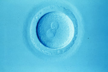
Oocytes mature differently in older women, in a process that could contribute to a decrease in fertility, a study from scientists in Barcelona, Spain has shown.
Scientists found that the activity of genes involved in chromosome segregation increased in oocytes progressively with age, while the activity of genes involved in mitochondrial metabolism decreased. Chromosome segregation is needed for cells to divide and multiply, and for RNA processing, while mitochondrial metabolism is essential for cell survival as mitochondria generate the energy needed for all cellular processes.
'Here we show that the final step of oocyte maturation itself might be negatively affected by age, which is critical for reproduction because [the oocyte] provides the material early embryos need to develop normally and survive,' Professor Bernhard Payer, co-author of the study, told Science Daily.
Researchers analysed 72 oocytes at different stages of maturity from 37 donors aged 18 to 43. They looked at the RNA molecules within each cell, known as the transcriptome, to see which genes show different levels of activity in oocytes from women of different ages. The findings were published in the journal Aging Cell.
Age-related changes to the transcriptome of oocytes were found to mostly occur in the final stage of development of the cell, when culturing in vitro. This finding reveals a potential pathway by which age could have an impact on the quality of oocytes, and why fertility is seen to decrease with age in women.
Further analysis revealed a number of genes that regulate the transcription of other genes could be behind these differences in the transcriptome of oocytes from women of different ages, and could be the focus of further research in this area. Transcription is the process of copying a segment of DNA into RNA. The transcribed segments then direct the synthesis of proteins.
The researchers also reported that higher body mass index (BMI) was found to impact oocyte development. Here however, the genetic activity differed in immature oocytes between women with different BMI. Thus it is likely BMI influences fertility through an alternate mechanism compared to the fertility decline caused by age. Women in the study mostly had a BMI that was normal or in the overweight category, with one donor obese and one donor underweight. As only one egg donor in the study was underweight, no conclusions could be drawn as to the effect of reduced BMI on egg quality.
The authors concluded that their work may serve as a resource for the development of future diagnostic tools to assess oocyte quality.


Leave a Reply
You must be logged in to post a comment.