The advent of PGD extended the scope of IVF beyond the treatment of infertility. PGD involves the removal of one or two cells from the early embryo to test for a specific genetic defect. It is predominantly used to prevent transmission of genetic defects arising from mutations in nuclear DNA, which encodes the characteristics we inherit from our parents. However, PGD can also be used to reduce the risk of transmitting mutations in mitochondrial DNA (mtDNA), which cause a range of debilitating and life-threatening diseases.
Mitochondrial DNA is contained in mitochondria (the cell's batteries), which are dispersed throughout the cell. Unlike nuclear DNA, which we inherit from both parents, mtDNA is inherited exclusively from our mothers. Mutations in mtDNA can either be present in all mitochondria or in only a proportion of mitochondria. In the latter case, known as heteroplasmy, the severity of disease is proportional to the ratio of mutated to non-mutated mtDNA.
Because of the manner in which mitochondria segregate to different cell types during embryonic development, a woman who is heteroplasmic for a given mutation, and who herself has a low mutation load, can produce eggs with varying levels of mutation. Consequently, some embryos may contain sufficiently high levels to cause serious disease in the child. By sampling a cell from the embryo it is possible to select those with the lowest mutation load, thereby reducing the risk of disease. However, PGD is not useful for homoplasmic women, whose entire complement of mtDNA carries the mutation. Likewise, the embryos of a heteroplasmic woman with a high mutation load are likely to also contain a high proportion of mutated mtDNA.
An alternative approach, currently being developed, is to rid the egg of affected mitochondria. As the egg contains hundreds of thousands of mitochondria, it is not practical to transplant the mitochondria themselves. However, it is possible to transplant the nuclear DNA into another egg. In theory, this approach could be used to create a reconstituted egg containing nuclear DNA from the affected woman and mtDNA from an unaffected donor. This would enable women with mutated mtDNA to have a genetically related child without transmitting mtDNA disease.
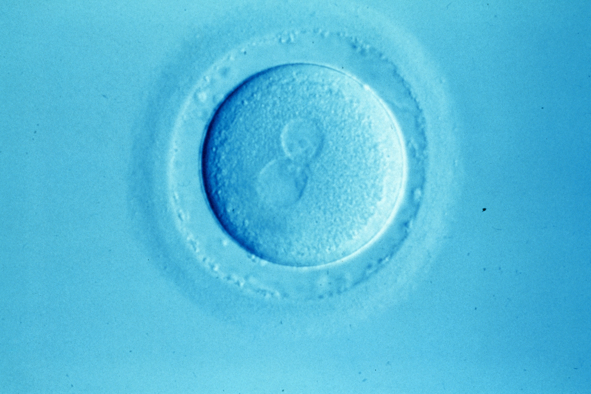
Two approaches to nuclear DNA transplantation are currently being explored. The first, which has been extensively tested in mice, is to transplant the nuclear DNA after the egg has been fertilised. At this stage the nuclear DNA from the egg and sperm is contained in two large structures, called the pronuclei, which are visible under the light microscope and can be transferred between fertilised eggs. The other approach is to transplant the nuclear DNA before fertilisation. At this stage the egg's nuclear DNA, poised to halve its content following sperm entry, is positioned within a structure known as the second meiotic spindle. Using specialised optics it is possible to visualise the spindle and to transplant it, together with the nuclear DNA, to another oocyte (egg). Pronuclear transfer has been found to be technically feasible in human eggs (1) and live births have been reported following spindle transfer in monkey oocytes (2). Either technique could, in principle, be used to dramatically reduce the risk of transmission of mtDNA disease.
Because the mitochondria are scattered throughout the egg, it is likely that a small proportion of mutated mtDNA will be transferred with the nuclear DNA. Thus, it is possible that the reconstituted egg will contain a small amount of mutated mtDNA. Studies in monkeys and humans indicate that this accounts for less than two percent of total mtDNA, whereas the threshold for disease is in the region of 60 percent mutation. Thus, on the basis of our current understanding, it is highly unlikely that a child born following pronuclear or spindle transfer will develop mtDNA disease. Nonetheless, ongoing research seeks to further reduce the level of mtDNA carryover.
The next big step is to determine which technique (pronuclear or spindle transfer) is likely to be the most efficient in terms of producing viable embryos. It will also be very important to determine whether embryos derived from reconstituted eggs differ from embryos produced by conventional IVF/ICSI procedures. While it was possible to perform proof of principle studies using abnormally fertilised human eggs, development of these embryos is impaired, making it impossible to perform meaningful analysis of safety and efficacy. It is therefore necessary to use eggs donated and fertilised specifically for the purpose of this research. Thus, the research, and indeed any future clinical application, will require a supply of donated eggs.
It is hoped that these studies will provide the evidence required for the regulators to make a decision on the suitability of nuclear DNA transplantation for clinical application. From a clinical embryology perspective, this is an enormously exciting development, which has the potential to further extend the scope of IVF-based technologies in preventing otherwise incurable diseases.


Leave a Reply
You must be logged in to post a comment.