Embryos have been grown in the lab for up to 25 days, the longest recorded in primates.
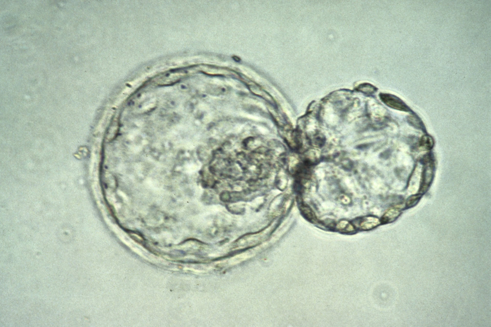
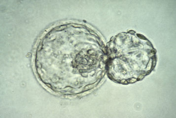
Until now, scientists have been unable to grow primate embryos in the lab for more than a couple of weeks, with the embryos not developing much beyond the blastocyst stage. Now, two separate research labs in China have shown how the embryos can be grown outside the uterus, for up to 25 days, enabling the observation of more advanced developmental milestones.
'... Our understanding of [the third and fourth weeks of gestation] is limited due to restricted access to in vivo embryos.' wrote one of the research teams in their paper published in Cell. 'This work... describes a system to study non-human primate embryogenesis,' wrote the other team in their paper, also published in Cell.
A major barrier to embryo research is the fact that, when grown in the uterus, watching the start of organ formation or the development of the nervous system, is limited. The two new studies describe how the growth of cynomolgus monkey embryos suspended in a gel-like growth medium allows them to develop in three dimensions (3D) as they would in the uterus.
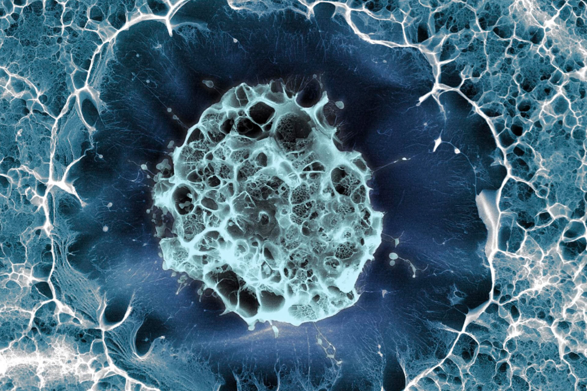
The first team, led by Professor Hongmei Wang of the State Key Laboratory of Stem Cell and Reproductive Biology in Beijing, had previously grown monkey embryos in the lab for up to 20 days. However, those embryos collapsed before reaching the early stages of nervous system and organ formation. Using their 3D method, the researchers were able to grow the embryos until they began to develop the three main layers that form different parts of the body – the endoderm, mesoderm, and ectoderm.
At the later stages of development, Professor Wang's team were able to observe the formation of heart muscle cells, early motor neurons and the neural plate – the beginnings of the brain and spine – which could be seen to bend into a tube as it formed the basis of the central nervous system.
The second team, led by Professor Tao Tan of the State Key Laboratory of Primate Biological Research in Yunnan, also found that the process of blood formation had begun in the yolk sacs.
'We were deeply impressed,' said Professor Tan, commenting that the blood cells 'are almost impossible to obtain during human embryonic development.'
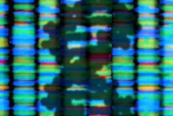
Analysis of gene expression in the embryos revealed the formation of distinct cell types, which would be seen in an embryo that develops within a uterus, as the stem cells divide and take on their new roles in the developing body.
'It's very impressive,' said Professor Magdalena Zernicka-Goetz from the University of Cambridge, who was not involved in the work. 'It's going to bring a lot of new insights'.
Despite their similarity to in-utero grown embryos, the lab-grown ones still show some differences, suggesting there is more to learn about what coordinates their development. Dr Naomi Moris from the Francis Crick Institute, London, who also was not involved in the study said: 'They still look a bit different to how we would expect [them] to look at these stages… There's definitely still scope for improvement.'
The researchers hope that their breakthrough in lab-grown embryos will enable new insights into the process of primate early development by allowing its observation in real-time, as well as paving the way for embryos to be grown in the lab for even longer.
Sources and References
Related Articles
Gradient-inducing proteins crucial for embryo development
Proteins that exist in the junctions between cells have been shown to help create signalling gradients crucial for cell differentiation in human embryo models...
Gene's role in embryonic organ development discovered
Malformations in organ development have been found to be caused by mutations of a gene expressed in early embryogenesis...
Human cells mimicking early embryogenesis generated
A type of embryonic cell has been generated from human stem cells for the first time, providing a method to study post-implantation development...
Stem cells discovered that resemble early human embryo cells
Human cells behaving like those at a crucial embryological milestone have been identified, enabling insights into early development...
Early stage of human embryo development seen for the first time
High-resolution single-cell gene expression analysis has been performed on a gastrulating human embryo, providing landmark insights...
Leave a Reply
You must be logged in to post a comment.