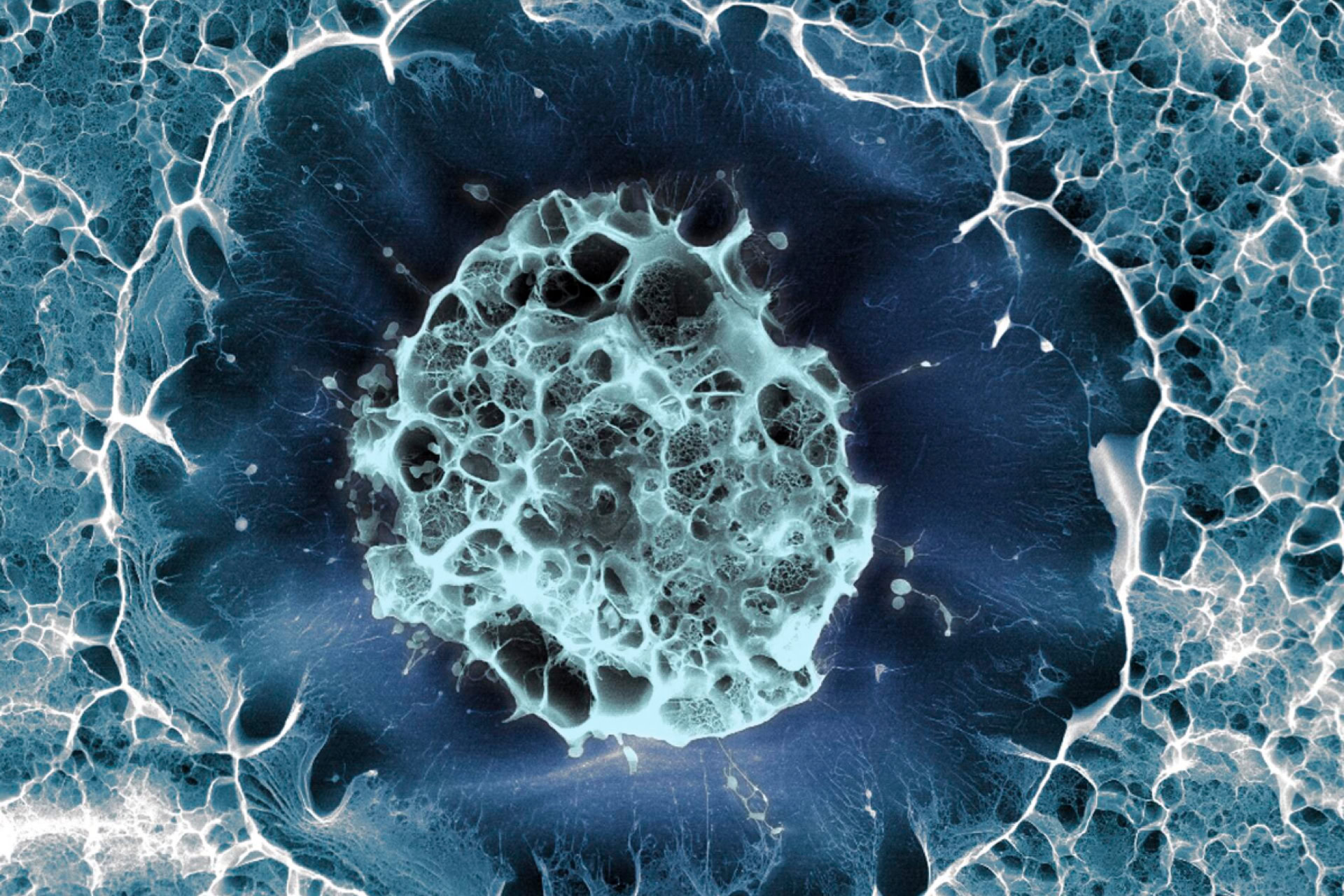
Stem cells collected from amniotic fluid during pregnancy can be grown into organoids, to study organ development and improve prenatal diagnosis of medical conditions.
Organoids are cellular structures that mimic human organs and can be used to study human development or model diseases. Often, these are generated from cells that have been reprogrammed into a stem-cell-like state, or from stem cells taken from tissue from aborted fetuses. This is the first published study that has taken stem cells shed by the fetus from the amniotic fluid, which were then grown to form organoids resembling small intestines, kidneys and lungs.
'This is the first time that we've been able to make a functional assessment of a child's congenital condition before birth, which is a huge step forward for prenatal medicine. Diagnosis is normally based on imaging such as ultrasound or MRI and genetic analyses,' said Professor Paolo de Coppi, senior author of the study, from University College London Great Ormond Street Institute of Child Health.
In the study, published in Nature Medicine, cells from the amniotic fluid were obtained during routine pregnancy procedures, either during amniocentesis (a diagnostic test for genetic or chromosomal conditions in which a needle is inserted into the womb) or through amniotic drainage (in which excess fluid is removed). In the UK and many other countries, samples of amniotic fluid are obtained for prenatal diagnosis of medical conditions.
The majority of the cells collected in this way were dead. However, a sufficient number of live cells were extracted to be grown in gel droplets. The researchers then sequenced RNA from the organoids to determine which tissues the cells came from, and were successfully able to grow lung, kidney and small intestine organoids. These organoids expressed similar genes and proteins to the relevant organs, as well as exhibiting functional similarities.
The researchers studied how such organoids could be used to study diseases, by growing organoids from stem cells collected from fetuses with congenital diaphragmatic hernia (CDH). This is a condition in which the diaphragm fails to develop, causing the intestine and liver to push into the chest and causing excess pressure on the lungs. The CDH organoids showed characteristic differences to those generated from healthy fetuses.
Sarah Norcross, director of PET (the Progress Educational Trust), said: 'Generating organoids during a pregnancy makes for a promising addition to established ways of studying early human development. While the study of cells and tissues obtained from terminations will remain vitally important to research, this new approach has the potential to offer different insights.'
The study collected cells from a relatively small number of pregnancies, a small sample, and the approach requires further validation. It is hoped that this approach to creating organoids may prove useful for purposes including disease screening and drug testing.
Sources and References
-
Stem cells collected in late pregnancy herald advances in prenatal medicine
-
Single-cell guided prenatal derivation of primary fetal epithelial organoids from human amniotic and tracheal fluids
-
Organoids grown from amniotic fluid could shed light on rare diseases
-
Organoids made from uterus fluid may help treat fetuses before birth
-
Scientists grow 'mini-organs' from cells shed by fetuses in womb
-
Scientists can help fetuses by growing tiny replicas of their organs
-
Organoids grown from late-stage foetuses offer boost for prenatal medicine
-
Organoids made from amniotic fluid will tell us how fetuses develop


Leave a Reply
You must be logged in to post a comment.