The energy required for our cells to function properly is mainly produced by mitochondria. Mitochondria are tiny structures within our cells, which contain their own DNA. The mitochondrial DNA (mtDNA) encodes a small number of the many proteins required to produce energy efficiently. Mutations in mtDNA cause a broad spectrum of diseases and degenerative disorders, which can be fatal.
In contrast to nuclear DNA, which we inherit from both parents, our mtDNA is inherited exclusively from our mothers. Thus, the mitochondria contained in the egg constitute the founder population for life. In cases where a woman carries a mix of mutated and non-mutated mtDNA, the inheritance of the mutation is complicated by the fact that the level of mutated mtDNA contained in her germ cells can vary widely. Consequently, a woman who herself carries a low mutation load is at risk of passing high levels of mutated mtDNA to her children.
In principle, it is possible to reduce the risk of transmitting mtDNA disease by using IVF-based techniques to uncouple the inheritance of mtDNA from nuclear DNA. By transplanting the nuclear DNA from the egg of an affected woman to that of a healthy donor from which the nuclear DNA has already been removed, it would be possible for an affected woman to have a genetically related child, with minimal risk of transmitting mtDNA disease.
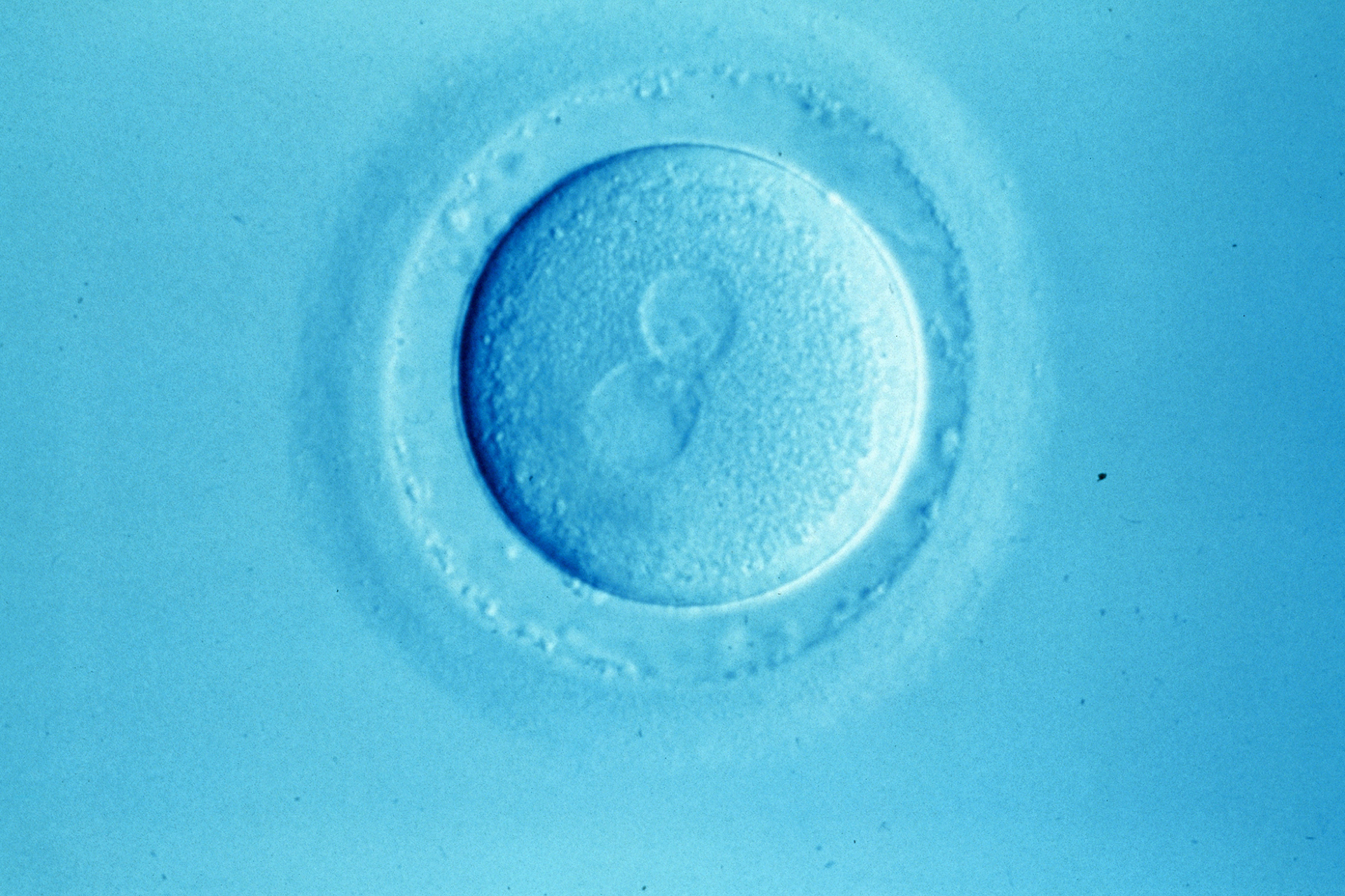
Transplantation of the nuclear DNA can be performed either before or after the egg is fertilised. Before fertilisation the egg's DNA, packaged into pairs of sister chromosomes, is positioned on a structure known as the meiotic spindle. Sperm entry triggers a series of biochemical changes in the egg, which cause the egg’s chromosomes to separate and move to opposite poles of the meiotic spindle. The chromosomes on the outermost pole are ejected from the egg in a small structure known as the polar body, while those on the inner spindle pole are retained in the egg. The chromosomes remaining in the egg then become enclosed in a large nucleus known as the female pronucleus. Around the same time the sperm chromosomes become enclosed in the male pronucleus. Normally fertilised eggs, therefore, contain two clearly visible pronuclei containing the nuclear DNA we inherit from both parents.
The technique of transplanting pronuclei between fertilised eggs (also known as zygotes) was pioneered in the mouse almost 30 years ago (1), and was found to be compatible with normal development over several generations of mice. More recently, this approach has been found to be feasible in human zygotes (2).
Transplantation of nuclear DNA between unfertilised eggs is a bit more complicated. It involves transplanting the meiotic spindle, which can only be visualised using specialised optics. Moreover, maintaining the correct positioning of chromosomes on the meiotic spindle is a biologically demanding task, which is susceptible to perturbation by changes in the egg's external environment. In this sense, compared with pronuclear transfer, the spindle transfer technique is more complex both from a technical and biological perspective.
Despite the complexities, a recent paper published by a US-based group reports that spindle transfer is also feasible in human eggs (3). This confirms previous findings from the same lab using rhesus monkey eggs (4). However, in contrast to their work on monkey eggs, these researchers found that abnormal fertilisation was prevalent in human eggs following spindle transfer. More than half of the eggs contained an abnormal number of pronuclei, which was largely due to the presence of an extra set of maternal chromosomes. The authors concluded that the manipulations required for spindle transplantation caused the egg's chromosomes to separate prematurely, and those that are normally lost to the polar body were retained in the egg.
What do these findings tell us? First, they tell us that human eggs are more sensitive to manipulation than those of rhesus monkey. Second, they tell us that the prevalence of abnormal fertilisation is likely to limit the efficacy of the spindle transfer technique, and that further work is required to overcome this problem. Importantly, the alternative technique of pronuclear transfer bypasses this hurdle by allowing the egg to separate its chromosomes and become packaged into pronuclei before the manipulations are performed.
Despite uncovering the problem with fertilisation, Tachibana and colleagues (2012) reported that when eggs fertilised normally following spindle transfer, the embryos developed well under laboratory conditions and gave rise to embryonic stem cell lines. Thus, while refinements are required, the study provides further encouraging evidence in support of the feasibility of IVF-based techniques in preventing transmission of mtDNA disease.
The next crucial step in progress towards the clinical application of these IVF-based techniques is to perform detailed comparisons between manipulated and unmanipulated embryos. This is essential to provide prospective patients with the evidence they require to make informed choices.
Sources and References
-
1) McGrath, J; Solter, D; 'Nuclear transplantation in the mouse embryo by microsurgery and cell fusion'
-
2) Craven, L. et al; 'Pronuclear transfer in human embryos to prevent transmission of mitochondrial DNA disease'
-
3) Tachibana et al; 'Towards germline gene therapy of inherited mitochondrial diseases'
-
4) Tachibana et al; 'Mitochondrial gene replacement in primate offspring and embryonic stem cells'


Leave a Reply
You must be logged in to post a comment.