



DNA in our cells is organised into units called chromosomes. Human embryos normally inherit 46 chromosomes: 23 from each parent. However, this process can sometimes go awry, resulting in embryos with missing or extra chromosome copies, a condition known as aneuploidy.
Having an abnormal number of chromosomes alters the dosage of hundreds of genes, causing many proteins to be produced in insufficient or excessive amounts, disrupting normal cell function. This often leads to pregnancy loss or, less commonly, to genetic conditions such as Down's syndrome.
PGT-A (pre-implantation genetic testing for aneuploidy) is a set of tests developed in the 1990s to assess the chromosome status of embryos during IVF treatment. These tests can be used by fertility clinics to help decide which embryos to transfer to patients. However, there are important nuances to assessing an embryo's genetic content that need to be considered.
For example, which chromosome – or chromosomes – are affected? Are there missing or additional chromosomes? And, crucially for this discussion, did the chromosomal abnormality originate in the gametes (egg or sperm) or did it arise during early embryo development?
The last point is particularly important. If an error occurs before fertilisation, the whole embryo will be aneuploid. If, on the other hand, the problem arises later in development, the embryo will contain a mixture of cells with normal and abnormal DNA content – a condition known as mosaicism.
Today, the most common version of PGT-A offered by fertility clinics involves taking a biopsy on day five of development. At this stage, the embryo is a spherical structure known as a blastocyst. It is comprised of a layer of outer cells that will become the placenta, and an inner cell mass that will give rise to the fetus, as well as tissues, including the yolk sac, which support embryo development before the placenta is fully established.
The biopsy removes approximately five cells from the outer placental precursor layer to determine their DNA content (nowadays typically using whole genome sequencing). There are three possible results following PGT-A:
- All cells tested have normal chromosome content.
- All cells are aneuploid.
- The biopsy is mosaic.
However, the results can be difficult to interpret, for several reasons.
Cell division errors are common during early embryo development, leading to a high prevalence of mosaicism. As a result, a PGT-A result based on five cells cannot be accurately extrapolated to the remaining 95 or so cells making up the blastocyst.
After cell division, the resulting daughter cells remain in close proximity to one another. This means that if an error occurred during a recent cell division, only the nearby cells will be abnormal. Consequently, the biopsy may produce a biased result because it samples a cluster of neighbouring cells from the same region of the embryo.
Sampling outer, placental-fated cells also does not indicate the chromosome status of the inner cells that will give rise to the fetus. This problem is compounded by the observation that placental cells are much more tolerant of chromosomal abnormalities than fetal cells, as demonstrated by reports of chromosomally normal babies whose pregnancies were supported by placentas with abnormal DNA content.
Moreover, analysis of whole blastocysts following PGT-A has shown that when outer cell biopsies exhibit low-to-medium levels of mosaicism (up to 50 percent aneuploidy), the chromosomal abnormalities extend to other parts of the embryo in only around one percent of cases. Transfer of these embryos, which have low-to-medium levels of mosaicism as assessed by PGT-A, has resulted in the births of babies with a normal chromosome number.
Despite this, there is reluctance to transfer embryos whose biopsies show high levels of aneuploidy (more than 50 percent). While such caution is understandable, it is important to note that some embryos initially characterised as fully aneuploid by PGT-A were subsequently shown to be mosaic. It could therefore be argued that patients should be given the choice of using such embryos, particularly if they have no other embryos available.
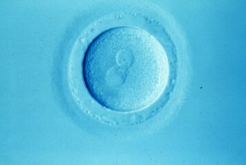
A different version of PGT-A, now used far less frequently, involves polar body biopsy. Polar bodies are formed during egg development as a way of eliminating half of the 46 chromosomes in preparation for fertilisation. A polar body can therefore be used as an indicator of an egg's chromosome status. Any embryos developing from aneuploid eggs will themselves be fully aneuploid, meaning polar body biopsy provides a way of identifying embryos with the highest proportions of aneuploid cells.
This is particularly relevant for older female patients, who are at increased risk of producing eggs with abnormal chromosome content. However, polar body biopsy also has downsides. These include providing no information about the chromosome status of the fertilising sperm and frequent failure to extract DNA of sufficient quality from the polar body to carry out the test.
Both versions of PGT-A require eggs or embryos to be removed from the controlled environment of the incubator and subjected to biopsy to obtain material for testing. While these procedures are compatible with successful live birth, there remains a lack of extensive studies examining their effect on embryo developmental potential and the long-term health outcomes of children born following these procedures.
In the UK, the Human Fertilisation and Embryology Authority gives PGT-A mixed ratings as an IVF add-on. It concludes that PGT-A can reduce the risk of miscarriage but may also decrease the chance of a live birth by the end of fertility treatment. PGT-A should not be confused with PGT-M or PGT-SR, embryo tests that, as the HFEA explains, can accurately confirm the presence or absence of specific disease-causing genetic variants.
While PGT-M and PGT-SR continue to hold promise as ways of reducing the risk of passing on genetic conditions to the next generation, PGT-A is inherently probabilistic and is therefore not sufficiently accurate to be used as a diagnostic tool.
Related Articles
Non-invasive PGT-A did not improve pregnancy rates
A non-invasive form of PGT-A did not improve pregnancy rates in a clinical trial, but did reduce early miscarriages...

PET Podcast: PGT-A as an IVF Add-On – 25 Years of Controversy
Bringing you PET events as a podcast. In the 40th episode of its podcast, PET discusses longstanding controversies surrounding PGT-A, with Dr Deborah Cohen, Professor Karen Sermon, Dr Justin Chu, James Lawford Davies and Professor Manuela Perrotta...


PGT-A as an IVF Add-On: 25 Years of Controversy
The second session of 'What Does Genomics Mean for Fertility Treatment?' – the 2025 Annual Conference of PET – explored controversies surrounding preimplantation genetic testing for aneuploidy...

Seeing the unseen: Live imaging of late-stage preimplantation human embryos
A novel method for visualising human blastocyst development – which has led to findings with implications for PGT-A – is described by Dr Ahmed Abdelbaki, a postdoctoral researcher in the Niakan Lab at the University of Cambridge...
PGT-A may overestimate impact of embryo abnormalities
A common IVF 'add-on' – preimplantation genetic testing for aneuploidy (PGT-A) – may overestimate the impact of embryo abnormalities, new research suggests...
An open letter to the HFEA about Choose a Fertility Clinic
This open letter to the UK fertility regulator has been written by fertility professionals at Guy's & St Thomas' NHS Foundation Trust, King's College London and the University of Southampton...
Class action lawsuits against genetic testing companies over PGT-A
Former IVF patients in the USA have filed class action lawsuits against several companies for misleading them about the efficacy of PGT-A...